1 UMN LESION Disrupts the balance of supraspinal inhibitory and - - PDF document

1
SMART_READER_LITE
LIVE PREVIEW

1 UMN LESION Disrupts the balance of supraspinal inhibitory and - - PDF document

UPPER An upper motor neuron lesion (also MOTOR known as pyramidal insufficiency ) is a lesion of the neural pathway above NEURON the anterior horn cell of the spinal cord LESION and/or motor nuclei of the cranial nerves. Corticospinal


slide-1
SLIDE 1

1

UPPER MOTOR NEURON LESION

  • An upper motor neuron lesion (also

known as pyramidal insufficiency) is a lesion of the neural pathway above the anterior horn cell of the spinal cord and/or motor nuclei of the cranial nerves.

  • Corticospinal and Corticobulbar

Tracts

  • This is in contrast to a lower motor

neuron lesion, which affects nerve fibers traveling from the anterior horn

  • f the spinal cord or the cranial motor

nuclei to the relevant muscle(s)
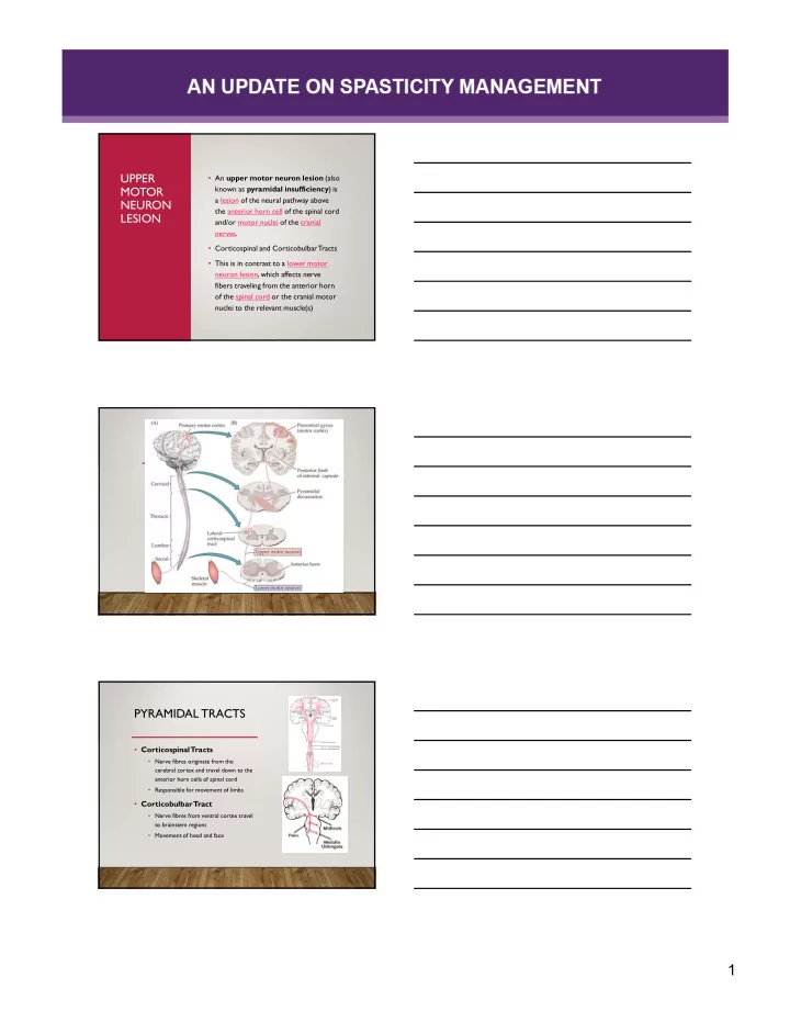
PYRAMIDAL TRACTS

  • Corticospinal

Tracts

  • Nerve fibres originate from the

cerebral cortex and travel down to the anterior horn cells of spinal cord

  • Responsible for movement of limbs
  • Corticobulbar

Tract

  • Nerve fibres from ventral cortex travel

to brainstem regions

  • Movement of head and face
slide-2
SLIDE 2

2

UMN LESION

  • Disrupts the balance of

supraspinal inhibitory and excitatory inputs directed to the spinal cord

  • Leading to a state of

disinhibition of the stretch reflex

  • = Hyperexcitability of

Stretch Reflex

MUSCLE HYPERTONIA

  • Extrinsic Hypertonia
  • Hyperexcitation of Stretch Reflex
  • = Spasticity, clonus, exaggerated tendon jerks, extensor spasm,

flexor withdrawal spasm, spastic dystonia

  • Intrinsic Hypertonia
  • Due to factors such as muscle contracture
  • Also known as nonreflex muscle hypertonia
  • Animal studies show that muscle immobilization at short lengths reduces

serial sarcomere number and increases the proportion of connective tissue in the muscle.

1.
  • J. C. Tabary, C. Tabary, C. Tardieu, G. Tardieu, and G. Goldspink, “Physiological and structural changes in the cat's soleus
muscle due to immobilization at different lengths by plaster casts,” Journal of Physiology, vol. 224, no. 1, pp. 231–244, 1972. 2.
  • T. A. H. Järvinen, L. Józsa, P. Kannus, T. L. N. Järvinen, and M. Järvinen, “Organization and distribution of intramuscular
connective tissue in normal and immobilized skeletal muscles,” Journal of Muscle Research and Cell Motility, vol. 23, no. 3,
  • pp. 245–254, 2002.

OTHER FORMS OF HYPERTONIA

  • RIGIDITY
  • Often seen with hypokinesia due to BG

disorders

  • does not depend on imposed speed and can

be elicited at very low speeds of passive movement

  • felt in both agonist and antagonist muscles

and in movements in both directions

  • Cogwheel rigidity and Leadpipe rigidity are

two types identified with Parkinson's disease

slide-3
SLIDE 3

3

OTHER FORMS OF HYPERTONIA

  • DYSTONIA
  • Patients experience muscle twisting, repetition, and abnormal

postures

  • Abnormal postures (i.e. cervical dystonia) can be present at rest or
  • nly brought on my certain actions (i.e. task specific dystonia or
  • ther limb dystonia)
  • Causes can be genetic, secondary to drugs or CNS trauma or

idiopathic

CONTRACTURE

  • A muscle contracture is a permanent

shortening of a muscle or joint = A Rigid Joint

  • Muscle or tendon too short for too long

Contracture

  • Spasticity and spastic dystonia can contribute

heavily to contracture development

  • Others causes
  • Ischemia (Volkmann’s)
  • Burns
  • Heterotopic Ossification
slide-4
SLIDE 4

4

slide-5
SLIDE 5

5

slide-6
SLIDE 6

6