SLIDE 1 5/26/2016 1
Joseph Rabban MD MPH Pathology Department University of California, San Francisco
Pelvic Serous Carcinoma: 2014 W.H.O. Update
Practical Implications for Pathologists
Outline of Talk
- Changes to 2014 WHO system for pelvic serous tumors
- High grade serous carcinoma versus low grade serous carcinoma
- New recommendations for assigning primary tumor origin
- Special issues with serous borderline tumors
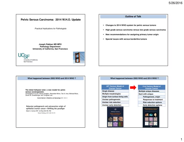
What happened between 2003 WHO and 2014 WHO ? What happened between 2003 WHO and 2014 WHO ?
20th Century Model of Ovarian Cancer Single disease Multiple morphologies Origin from surface lining cells Unclear pathogenesis Unclear risk reduction Unclear early detection 21st Century Model of Ovarian Cancer Multiple unique diseases Each with unique: Pathogenesis, origin Responses to treatment Risk reduction options Early detection options
SLIDE 2
5/26/2016 2
Ovarian Carcinomas in 2014 WHO High grade serous carcinoma (HGSC) Low grade serous carcinoma (LGSC) Clear cell carcinoma Endometrioid adenocarcinoma Mucinous carcinoma 3 Major Changes to Pelvic Serous Tumors in 2014 WHO
2003 2014 1 Tumor Class Surface epithelial – stromal tumors Epithelial tumors
Rationale:
New Evidence Tumor origin is fallopian tube mucosa (either in tube as STIC or in ovary as epithelial inclusion cyst) Implications Fallopian tube is target for risk reduction, early detection P53
Tubal HGSC (STIC) Ovarian HGSC Proposed Tubal Origins of Ovarian HGSC versus LGSC Benign tubal mucosa Inclusion gland
P53
Ovarian HGSC Proposed Tubal Origins of Ovarian HGSC versus LGSC
SLIDE 3 5/26/2016 3
Benign tubal mucosa Inclusion gland Ovarian Serous Borderline Tumor
BRAF KRAS MAPK pathway
Ovarian LGSC Proposed Tubal Origins of Ovarian HGSC versus LGSC Inclusion gland Ovarian Serous Borderline Tumor
BRAF KRAS MAPK pathway
Ovarian LGSC
p53
Tubal HGSC (STIC)
p53
Ovarian HGSC
spread benign
Proposed Tubal Origins of Ovarian HGSC versus LGSC 3 Major Changes to Pelvic Serous Tumors in 2014 WHO
2003 2014 1 Tumor Class Surface epithelial – stromal tumors Epithelial tumors 2 Serous Carcinoma One tumor, grade 1,2,3 Two tumors: HGSC, LGSC New Evidence Distinct behavior, response to therapy, genetics, pathogenesis, origin Implications Distinct surgical planning, adjuvant therapy, genetic counseling, risk reduction
Rationale: 3 Major Changes to Pelvic Serous Tumors in 2014 WHO
2003 2014 1 Tumor Class Surface epithelial – stromal tumors Epithelial tumors 2 Serous Carcinoma One tumor, grade 1,2,3 Two tumors: HGSC, LGSC 3 Transitional cell carcinoma Unique tumor
variant of HGSC New Evidence “TCC” has same properties as HGSC Implications Diagnosis of HGSC includes many different patterns beyond papillary (TCC, endometrioid, solid)
Rationale:
SLIDE 4 5/26/2016 4
Outline of Talk
- Changes to 2014 WHO system for pelvic serous tumors
- High grade serous carcinoma versus low grade serous carcinoma
- New recommendations for assigning primary tumor origin
- Special issues with serous borderline tumors
High Grade Serous Carcinoma Low Grade Serous Carcinoma HGSC LGSC LGSC: Clinico-pathologic Features Incidence ~5 % of all ovarian cancers Age Average 4th to 5th decade Risk factors No traditional HGSC risk factors 20% risk if prior history of advanced stage serous borderline tumor ~ risk if prior ovulation induction for fertility Hereditary ? No major syndrome known Stage >75% are advanced stage at diagnosis Median survival ~80-96 months Time to recurrence ~33 months if optimal cytoreduction ~14 months if suboptimal cytoreduction
Romero 2013 Gyn Oncol Fader 2013 Ob Gynecol
HGSC LGSC BRCA hereditary syndrome ~25 % No P53 mutation Yes No BRAF KRAS MAPK pathway defects No Common Precursor lesion STIC Borderline tumor Platinum chemotherapy sensitive Yes Uncommon PARP inhibitor sensitive Yes Unlikely Neoadjuvant chemotherapy candidate Yes No MAPK pathway inhibitor candidate No Yes
SLIDE 5 5/26/2016 5
HGSC
Papillary Micro-papillary Solid Pseudo-endometrioid (cribriform) Transitional cell carcinoma-like High nucleus-cytoplasm ratio Pleomorphism Nuclear hyperchromasia Brisk / atypical mitoses Macro-nucleoli
HGSC: Papillary pattern HGSC: Papillary pattern
Pleomorphism, high N/C ratio, brisk mitoses
HGSC: Transitional cell cancer-like pattern
SLIDE 6 5/26/2016 6
HGSC: Transitional cell cancer-like pattern HGSC: Pseudo-endometrioid pattern HGSC: Pseudo-endometrioid pattern HGSC
Mullerian origin + PAX8, CK7 Extra-uterine serous differentiation + WT1 High grade serous carcinoma Aberrant p53, p16
SLIDE 7
5/26/2016 7
Normal p53 gene p53 IHC stain result p53 and Pelvic HGSC Weak patchy staining (“Normal” / Wild type) p53 gene mutation p53 IHC stain result p53 and Pelvic HGSC
~80% ~20%
Diffuse, strong staining Completely negative
Pattern of p53 IHC staining Stain Interpretation Diagnosis Strong/diffuse Aberrant p53 HGSC Completely negative Weak/patchy Wild type p53 (normal) not HGSC *
p53 Stain Interpretation in Pelvic Serous Carcinoma
Wild type p53 Aberrant p53 Aberrant p53 HGSC Check for internal control HGSC not HGSC*
p53 Stain Interpretation in Pelvic Serous Carcinoma
SLIDE 8 5/26/2016 8
Diffuse / strong = Aberrant p16 Patchy or negative = Wild-type p16
p16 Stain Interpretation in Pelvic Serous Carcinoma Use both p53 and p16 stains for HGSC
Cannot use p53 alone if result is wild type 4% of HGSC are wild type p53 / aberrant p16 Always do both stains together
LGSC
Papillary branching Micro-papillary Bud-like Macro-papillary Cribriform Uniform, monotonous Focal moderate atypia (<3:1 variable size) Mitoses < 12 / 10 hpf Atypical mitoses uncommon
MD Anderson Criteria for LGSC
Grading System # of Grading Tiers FIGO 3 Shimizu-Silverberg 3 MD Anderson 2
SLIDE 9
5/26/2016 9
FIGO Universal Grading of Pelvic Cancer (any type)
Grade Solid Architecture 1 <6 % 2 6 % – 50% 3 > 50%
Shimizu-Silverberg Universal Grading of Ovarian Cancer (any type)
Points Architecture Atypia Mitoses /10 hpf 1 Glandular Mild 0-9 2 Papillary Moderate 10-24 3 Solid Severe >24 Total Points Overall Grade 3 – 5 1 6, 7 2 8, 9 3
MD Anderson Classification of Ovarian Serous Carcinoma Classification Atypia Mitoses / 10 hpf LGSC Mild to Moderate < 12 HGSC Marked 12 or more Nuclei Mild-Moderate Atypia Marked Atypia Nuclear appearance Uniform Pleomorphic Nuclear size/shape < 3 : 1 variability >3 : 1 variability Nucleoli None to small Macro Chromatin Evenly dispersed Coarse MD Anderson Classification of Ovarian Serous Carcinoma Classification Associated Borderline Tumor Progression Free Survival LGSC 60 % of cases longer HGSC 2 % of cases shorter Additional Advantages of 2-tier grading: More predictive than other grading systems Reproducible (Malpica 2007 AJSP) Eventually was validated by:
p53/p16 immunophenotype Molecular pathogenesis Response to therapy Association with inherited mutations
Used by 2014 W.H.O.
SLIDE 10 5/26/2016 10
LGSC with Serous Borderline Tumor LGSC: Micropapillary Buds (avascular) LGSC: Micropapillary Buds (avascular)
<3:1 varying size/shape No mitoses No macronucleoli
LGSC: Micropapillary Buds (avascular)
<3:1 varying size/shape No mitoses No macronucleoli
SLIDE 11 5/26/2016 11
LGSC: Micropapillary Buds (avascular)
<3:1 varying size/shape No mitoses No macronucleoli
LGSC: Papillary Branching (vascularized) LGSC: Papillary Branching (vascularized)
<3:1 varying size/shape No mitoses No macronucleoli
LGSC: Papillary Branching + Buds
SLIDE 12 5/26/2016 12
LGSC: Papillary Branching + Buds LGSC: Papillary Branching + Buds
<3:1 varying size/shape No mitoses No macronucleoli
LGSC: Branching + Cribriform Pattern LGSC: Branching + Cribriform Pattern
SLIDE 13
5/26/2016 13
LGSC: Cribriform Pattern LGSC: Columnar cells LGSC: Columnar cells Positive WT1 Excludes Endometrioid Tumor LGSC with Necrosis
SLIDE 14 5/26/2016 14
Most LGSC Present at Advanced Stage Pelvic Lymph Node Metastasis Most LGSC Present at Advanced Stage Omental Involvement Most LGSC Present at Advanced Stage Lung Metastasis
LGSC
Mullerian origin + PAX8, CK7 Extra-uterine serous differentiation + WT1 Low grade serous carcinoma Wild type p53, p16
SLIDE 15 5/26/2016 15
Wild type p53 in LGSC LGSC Wild type p53 Wild type p16 Wild Type p53 and p16 staining
89 % Sensitivity for LGSC 93 % Specificity 98 % Negative predictive value Aberrant results exclude LGSC Diagnostic Challenges Distinguishing LGSC versus HGSC Only moderate nuclear atypia Abundant cytoplasm Architecture of borderline tumor
Micropapillary architecture LGSC with: HGSC with: Focal mitotic activity Notable moderate atypia Rare severe atypia
SLIDE 16 5/26/2016 16
Diagnostic Challenges Distinguishing LGSC versus HGSC Only moderate nuclear atypia Abundant cytoplasm Architecture of borderline tumor
Micropapillary architecture LGSC with: HGSC with: Focal mitotic activity Notable moderate atypia Rare severe atypia p53, p16 Immuno Staining Resolve by: HGSC with only moderate atypia + rare mitoses HGSC with only moderate atypia + abundant cytoplasm HGSC with only moderate atypia + abundant cytoplasm
SLIDE 17
5/26/2016 17
Aberrant p53 Aberrant p16
HGSC with only moderate atypia + abundant cytoplasm HGSC with uniform micropapillary architecture HGSC with uniform micropapillary architecture HGSC with uniform micropapillary architecture P53
SLIDE 18 5/26/2016 18
HGSC mimicking Serous Borderline Tumor (conventional type) HGSC mimicking Serous Borderline Tumor (micropapillary type) HGSC mimicking Serous Borderline Tumor (micropapillary type)
Severe Atypia High Mitotic rate
HGSC mimicking Serous Borderline Tumor (micropapillary type)
SLIDE 19 5/26/2016 19
HGSC mimicking Serous Cystadenofibroma HGSC mimicking Serous Cystadenofibroma HGSC mimicking Serous Cystadenofibroma
Severe Atypia High Mitotic rate
LGSC has same survival as HGSC
Ali et al. 2013 Int J Gyn Path
SLIDE 20 5/26/2016 20
Why should pathologists distinguish HGSC vs LGSC ? Management Reasons to Distinguish HGSC vs LGSC
HGSC LGSC Consider Neoadjuvant chemotherapy ? Yes No Consider PARP inhibitor drug trials ? Yes No Consider MEK inhibitor drug trials ? No Yes Consider hormonal therapy ? No Yes Consider genetic counseling referral for risk of hereditary syndrome ? Yes Low priority
Neoadjuvant Chemotherapy for Advanced Stage Ovarian Cancer Same survival as primary surgery But:
- Decreased Morbidity
- Decreased Mortality
- Decreased Cost
- Informs About Response of
Selected Chemotherapy Agents
SLIDE 21 5/26/2016 21
LGSC is Often Resistant to Platinum Chemotherapy
Similar Findings: Schmeler 2008 Gynecol Oncol Gershenson 2009 Gynecol Oncol Schmeler 2011 Gynecol Oncol Ali 2013 Int J Gynecol Pathol Gershenson 2006 Obstet Gynecol
~ 50 % have residual disease after first round LGSC is Often Resistant to Neoadjuvant Chemotherapy Only ~ 5 % have any response
- Serous carcinoma, gynecologic origin
- Ovarian serous carcinoma
Clinically Advanced Stage Pelvic Cancer Biopsy of Omental Tumor or Paracentesis of Malignant Ascites Mullerian Adenocarcinoma (positive PAX8, WT1)
LGSC is Second Most Common Tumor Type In Advanced Stage Ovarian Cancer
Sub-Type % of All Advanced Stage Cases
HGSC 87.7 % LGSC 5.3 % Clear cell 4.5 % Endometrioid 2.5 % Mucinous 1.2%
Kobel et al. 2010 IJGP
SLIDE 22 5/26/2016 22
Neoadjuvant Chemo Tx
Primary Surgery Then Adjuvant Chemo Tx
Clinically Advanced Stage Pelvic Cancer Biopsy of Omental Tumor or Paracentesis of Malignant Ascites HGSC LGSC
Primary Surgery Then Adjuvant Chemo Tx
Diagnostic Challenges Distinguishing LGSC versus HGSC Morphology and immunostaining criteria not studied in : Core biopsy / FNA of peritoneal disease Cytology of malignant ascites
Management Reasons to Distinguish HGSC vs LGSC
HGSC LGSC Consider Neoadjuvant chemotherapy ? Yes No Consider PARP inhibitor drug trials ? Yes No Consider MEK inhibitor drug trials ? No Yes Consider hormonal therapy ? No Yes Consider genetic counseling referral for risk of hereditary syndrome ? Yes Low priority
Targeted Drugs in Clinical Trials for Pelvic Serous Carcinoma For HGSC (BRCA mutation): Targeted inhibitors of PARP1 protein For LGSC: Targeted inhibitor of MEK protein Olaparib Veliparib Rucaparib Selumetinib
SLIDE 23
5/26/2016 23
Normal DNA Repair Mechanisms Double Strand Breaks: Repair by BRCA1 / 2 proteins Single Strand Breaks: Repair by PARP1 protein If BRCA mutation, PARP1 “permits” Cells to Still Proliferate Double Strand Breaks: Repair by BRCA1 / 2 proteins Single Strand Breaks: Repair by PARP1 protein Alteration #1: Germline mutation BRCA1 or 2 Targeted Cell Death Occurs if PARP1 Inhibitor plus BRCA1 or 2 Double Strand Breaks: Repair by BRCA1 / 2 proteins Single Strand Breaks: Repair by PARP1 protein Alteration #1: Germline mutation BRCA1 or 2 Alteration #2: PARP1 Inhibitor Targeted Drug Targeted Drugs in Clinical Trials for Pelvic Serous Carcinoma For HGSC (BRCA mutation): Targeted inhibitors of PARP1 protein Olaparib Veliparib Rucaparib
SLIDE 24 5/26/2016 24
Management Reasons to Distinguish HGSC vs LGSC
HGSC LGSC Consider Neoadjuvant chemotherapy ? Yes No Consider PARP inhibitor drug trials ? Yes No Consider MEK inhibitor drug trials ? No Yes Consider hormonal therapy ? No Yes Consider genetic counseling referral for risk of hereditary syndrome ? Yes Low priority
Normal MAPK Cell Signaling Cascade Drives Proliferation
www.cancer.gov
TUMOR Mutations in MAPK Cell Signaling Cascade Promote Tumor Growth TUMOR MEK Inhibitor Drugs Shut Down Tumor Cell Proliferation MEK Inhibitor Drug
SLIDE 25 5/26/2016 25
Targeted Drugs in Clinical Trials for Pelvic Serous Carcinoma For LGSC: Targeted inhibitors of MEK protein in trials Selumetinib Trametinib
Management Reasons to Distinguish HGSC vs LGSC
HGSC LGSC Consider Neoadjuvant chemotherapy ? Yes No Consider PARP inhibitor drug trials ? Yes No Consider MEK inhibitor drug trials ? No Yes Consider hormonal therapy ? No Yes Consider genetic counseling referral for risk of hereditary syndrome ? Yes Low priority
Rationale for Hormonal Therapy in LGSC
Wong 2007 Int J Gynecol Pathol
LGSC HGSC Estrogen Receptors 58 % 27 % Progesterone Receptors 43 % 17 % Some survival benefit for LGSC with hormonal + chemo therapy
Gershenson 2012 Gyn Oncol Schlumbrecht 2011 Cancer
Management Reasons to Distinguish HGSC vs LGSC
HGSC LGSC Consider Neoadjuvant chemotherapy ? Yes No Consider PARP inhibitor drug trials ? Yes No Consider MEK inhibitor drug trials ? No Yes Consider hormonal therapy ? No Yes Consider genetic counseling referral for risk of hereditary syndrome ? Yes Low priority
SLIDE 26 5/26/2016 26
Genetic mutations can be found in up to 1/4 of all pelvic cancers Fanconi Anemia – BRCA Pathway Genes Mismatch Repair Genes TP53
- BRCA1, BRCA2
- RAD51C, RAD51D
- BRIP1
- BARD1
- CHEK2
- MRE11A
- NBN
- PALB2
- RAD50
- MLH1, MSH2, PMS2, MSH6
~80%
Walsh et al, 2011 PNAS
Unselected Pelvic Cancers
Most LGSC is not due to inherited gene mutation
No significant rate of germline mutations
No significant rate of “cancer pedigree” by personal/family history
Walsh 2011 PNAS Fujiwara 2012 AJSP Lakhani 2004 Clin Can Res Mavaddat 2011 Canc Epid Biom Prev Norquist 2013 Gyn Oncol Vineyard 2011 Gyn Onc
Management Reasons to Distinguish HGSC vs LGSC
HGSC LGSC Consider Neoadjuvant chemotherapy ? Yes No Consider PARP inhibitor drug trials ? Yes No Consider MEK inhibitor drug trials ? No Yes Consider hormonal therapy ? No Yes Consider genetic counseling referral for risk of hereditary syndrome ? Yes Low priority
Outline of Talk
- Changes to 2014 WHO system for pelvic serous tumors
- High grade serous carcinoma versus low grade serous carcinoma
- New recommendations for assigning primary tumor origin
- Special issues with serous borderline tumors
SLIDE 27 5/26/2016 27
How do we assign origin of advanced stage HGSC
- Traditional approaches:
- Problems
Any ovarian involvement = ovarian origin Dominant-mass = primary origin No consensus or standardization Observer variation Problematic in neoadjuvant treated cases Biologic validity is untested Does not address the new paradigm of fallopian tube findings How do we assign origin of advanced stage HGSC
Fallopian tube STIC is the earliest stage of HGSC
Setting % with STIC Incidental salpingectomy in general population < 1 % Prophylactic salpingectomy in BRCA mutation carrier < 8% Salpingectomy in stage I ovarian HGSC ~ 100 % Salpingectomy in advanced stage HGSC ~ 60 %
How do we assign origin of advanced stage HGSC
- New proposal from international consensus group:
How do we assign origin of advanced stage HGSC
- New proposal from international consensus group:
Use the presence of STIC to define the tube as primary origin
SLIDE 28 5/26/2016 28
How do we assign origin of advanced stage HGSC
- New proposal from international consensus group:
Use the presence of STIC to define the tube as primary origin
Pelvic HGSC % of all cases Primary fallopian tube cancer 83 % Primary ovarian cancer 17 % Primary peritoneal cancer 0 %
Singh 2015 Histopath
How do we assign origin of advanced stage HGSC
- New proposal from international consensus group:
- Advantages
Use the presence of STIC to define the tube as primary origin Simple rules Reproducible Applicable in neoadjuvant treated cases In sync with new paradigm of pathogenesis How do we assign origin of advanced stage HGSC
- New proposal from international consensus group:
- Unresolved issue: Uterine HGSC
Use the presence of STIC to define the tube as primary origin Primary uterine HGSC WT1 negative Primary extra-uterine HGSC WT1 positive Diagnostic Criteria for STIC
- Gross specimen management
- Morphologic criteria
- Immunohistochemical criteria
- Diagnostic pitfalls
SLIDE 29 5/26/2016 29
2 millimeter
STIC in Microscopic Process Tubes via SEE-FIM Protocol Fix in Formalin Several Hours Slice Fimbriae Parallel to Plicae 2-3 mm Intervals Embed Entire Fimbriae Entire tube if RRSO
Process Tubes via SEE-FIM Protocol Entire Fimbriae: Ampullary Portion of Tube 1-2 cassettes usually 1 cassette Automatic deeper levels not necessary if slices are 2-3 mm Diagnosis of STIC Morphology Architecture Cytology Immunophenotype Aberrant p53 Increased Ki-67 (MIB-1)
SLIDE 30 5/26/2016 30
Diagnosis of STIC Morphology Architecture Cytology
Crowding, tufting, piling up, stratification Loss of polarity Loss of cilia Loss of peg cells High N/C ratio Moderate / severe nuclear atypia Mitoses +/- Macronucleoli
Normal Fallopian Tube Mucosa STIC STIC
SLIDE 31
5/26/2016 31
STIC STIC Diagnostic Criteria Do Not Depend on BRCA mutation status Morphology Suspicious But Not Diagnostic of STIC Morphology Suspicious But Not Diagnostic of STIC
SLIDE 32
5/26/2016 32
Diagnosis of STIC Morphology Architecture Cytology Immunophenotype Aberrant p53 Increased Ki-67 (MIB-1) Other markers, not as well studied: p16, HMGA2, Stathmin Diagnosis of STIC p53 Stain Pattern Interpretation Strong + diffuse Aberrant Completely absent Aberrant Variable strength Normal (wild type) Ki-67 Stain Pattern Interpretation Same as adjacent normal mucosa Normal Higher than adjacent normal mucosa (>10%) Increased Aberrant p53 in STIC Aberrant P53 Aberrant Ki 67 STIC
SLIDE 33 5/26/2016 33
Aberrant P53 Aberrant Ki 67 STIC STIC Aberrant p53 Aberrant Ki67 Aberrant p16 Both Morphology and Immunostains Should Be Concordant for STIC
Vang IJGP 2012; 31: 243
Abnormal proliferations that fall short of criteria for STIC
SLIDE 34 5/26/2016 34
Abnormal proliferations that fall short of criteria for STIC Outcome based evidence of clinical significance is lacking Prone to observer variation No consensus on nomenclature No guidelines for management
Current UCSF approach is observation only
Serous tubal intraepithelial lesion Tubal intraepithelial lesion in transition Atypical mucosal proliferation
Alterations that are not clinically significant to report Secretory cell outgrowth (SCOUT) Alterations that are not clinically significant to report Secretory cell outgrowth (SCOUT) p53 signature (normal morphology but p53 aberrant) Alterations that are likely within normal spectrum of benign tubes “hyperplasia” without atypia (no formal terminology)
SLIDE 35 5/26/2016 35
Alterations that are likely within normal spectrum of benign tubes “hobnail” growth without atypia (no formal terminology)
Differential Diagnosis of STIC Tangential Sectioning Artifact Metaplasias (transitional cell, mucinous) Inflammatory Reactions Mucosal Endometriosis Mucosal Adenofibroma Submucosal Adenomatoid Tumor Metastatic Cancer Outline of Talk
- Changes to 2014 WHO system for pelvic serous tumors
- High grade serous carcinoma versus low grade serous carcinoma
- New recommendations for assigning primary tumor origin
- Special issues with serous borderline tumors
- Chemotherapy indications
LGSC in ovarian SBT LGSC in extra-ovarian sites
Special Issues with Serous Borderline Tumors (SBT)
SLIDE 36 5/26/2016 36
- Chemotherapy indications
- LGSC in ovarian SBT
LGSC in ovarian SBT LGSC in extra-ovarian sites Stromal invasive criteria (> 5 mm) Expansile invasion criteria
Special Issues with Serous Borderline Tumors (SBT) LGSC in Ovarian Serous Borderline Tumor > 5 mm span of stromal invasion LGSC versus Micropapillary Type SBT Special Issues with Serous Borderline Tumors (SBT)
- Chemotherapy indications
- LGSC in extra-ovarian sites (so-called “invasive” implants)
LGSC in ovarian SBT LGSC in extra-ovarian sites Destructive infiltrative growth into underlying tissue Solid nests / micropapillae within clefts in stroma
SLIDE 37 5/26/2016 37
LGSC / “Invasive Implant” in Omentum LGSC / “Invasive Implant” in Omentum
- Most SBT exhibit hierarchical branching growth
Special Issues with Serous Borderline Tumors (SBT)
- Micropapillary variant associated with extra-ovarian LGSC
Special Issues with Serous Borderline Tumors (SBT)
SLIDE 38 5/26/2016 38
- Micropapillary variant associated with extra-ovarian LGSC
Non-hierarchical branching Broad papillae with finger-like projections Broad papillae lined by cribriform proliferation > 5 mm span in conventional type SBT
Valuable to report at Frozen Section diagnosis in order to assist surgeon in considering staging procedure Special Issues with Serous Borderline Tumors (SBT)
Outline of Talk
- Changes to 2014 WHO system for pelvic serous tumors
- High grade serous carcinoma versus low grade serous carcinoma
- New recommendations for assigning primary tumor origin
- Special issues with serous borderline tumors