
SLIDE 1
MID 13
Anaerobes
Michael Yin, MD MS
Definitions
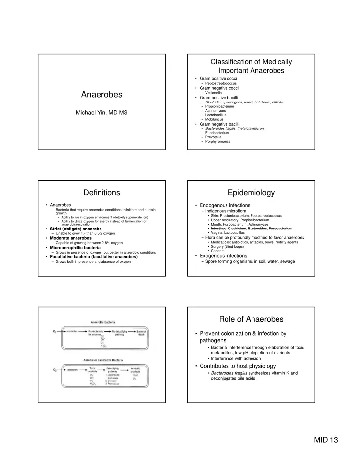
- Anaerobes
– Bacteria that require anaerobic conditions to initiate and sustain growth
- Ability to live in oxygen environment (detoxify superoxide ion)
- Ability to utilize oxygen for energy instead of fermentation or
anaerobic respiration
- Strict (obligate) anaerobe
- Strict (obligate) anaerobe
– Unable to grow if > than 0.5% oxygen
- Moderate anaerobes
– Capable of growing between 2-8% oxygen
- Microaerophillic bacteria
– Grows in presence of oxygen, but better in anaerobic conditions
- Facultative bacteria (facultative anaerobes)
– Grows both in presence and absence of oxygen
Classification of Medically Important Anaerobes
- Gram positive cocci
– Peptostreptococcus
- Gram negative cocci
– Veillonella
- Gram positive bacilli
Clostridium perfringens tetani botulinum difficile – Clostridium perfringens, tetani, botulinum, difficile – Propionibacterium – Actinomyces – Lactobacillus – Mobiluncus
- Gram negative bacilli
– Bacteroides fragilis, thetaiotaomicron – Fusobacterium – Prevotella – Porphyromonas
Epidemiology
- Endogenous infections
– Indigenous microflora
- Skin: Propionibacterium, Peptostreptococcus
- Upper respiratory: Propionibacterium
- Mouth: Fusobacterium, Actinomyces
- Intestines: Clostridium Bacteroides Fusobacterium
- Intestines: Clostridium, Bacteroides, Fusobacterium
- Vagina: Lactobacillus
– Flora can be profoundly modified to favor anaerobes
- Medications: antibiotics, antacids, bowel motility agents
- Surgery (blind loops)
- Cancers
- Exogenous infections
– Spore forming organisms in soil, water, sewage
Role of Anaerobes
- Prevent colonization & infection by
pathogens
- Bacterial interference through elaboration of toxic
metabolites, low pH, depletion of nutrients
- Interference with adhesion
- Contributes to host physiology
- Bacteroides fragilis synthesizes vitamin K and