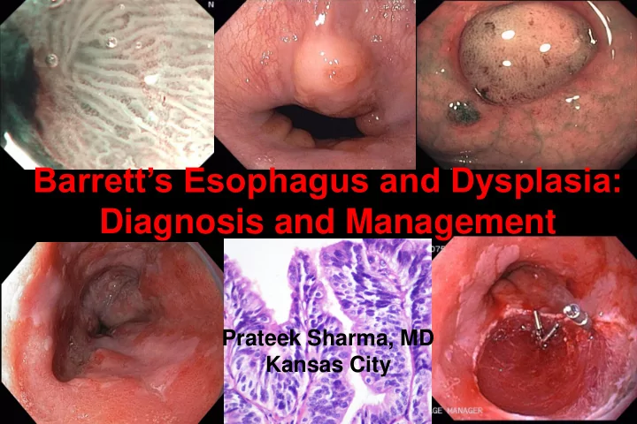
SLIDE 1
Barrett’s Esophagus and Dysplasia: Diagnosis and Management
Prateek Sharma, MD Kansas City
SLIDE 2 Barrett’s associated adenocarcinoma
squamous
Barrett’s
SLIDE 3 Rising Incidence of Esophageal Adenocarcinoma
Rate per 1,000,000 1975 2000 1980 1985 1990 1995 Pohl H et al, J Natl Cancer Inst 2005 5 10 15 20 25 30 35 Adenocarcinoma Squamous Cell Carcinoma Not otherwise specified
SLIDE 4
Intestinal Metaplasia
Barrett’s Esophagus
Columnar lined esophagus
SLIDE 5
SLIDE 6
Endoscopic recognition of the columnar lined esophagus
SLIDE 7
Long Barrett’s Short Barrett’s Ultra short Barrett’s Microscopic Barrett’s Invisible Barrett’s Terminology Issues
SLIDE 8 Prague C & M Criteria
Circumference and Maximum extent
Barrett’s, distal 2 cm circumferential and proximal 3 cm in form
Barrett’s: C2M5
C2 M5
Sharma P, Dent J, Armstrong D et al, Gastroenterology 2006
SLIDE 9
Progression of Barrett’s Esophagus
SLIDE 10 Dysplasia and cancer in BE patients: absolute risk
6.7 3 7.3 0.5 0.9 4.3 1 2 3 4 5 6 7 8 Cancers HGD LGD %
Prevalence
(n=1376)
Incidence
(n=618) Sharma et al, Clin Gastro Hepatol 2006
SLIDE 11
Endoscopic Therapy for Esophageal Neoplasia
Early Detection Accurate Staging Effective Treatment
SLIDE 12
Narrow Band Imaging (NBI)
Conventional imaging NBI
SLIDE 13
SLIDE 14
Field of view: 500x500µm Range: 0-250µm Lateral resolution: <1µm
Technique of Endomicroscopy
SLIDE 15
Endoscopic Therapy for Esophageal Neoplasia
Early Detection Accurate Staging Effective Treatment
SLIDE 16 EMR versus EUS
Baseline Diagnosis EMR Diagnosis
- 48 patients underwent EUS
- Invasion confirmed in 8 (7 at surgery)
Overall accuracy of EUS for staging 85% 1 over-staged, 6 under-staged
Larghi A et al, Gastrointest Endosc 2005
HGD (n=25) EUS: no cancer Cancers (n=15) EUS: intra mucosal 24% invasive cancer 40% invasive cancer
SLIDE 17
SLIDE 18
Endoscopic Therapy for Esophageal Neoplasia
Early Detection Accurate Staging Effective Treatment
SLIDE 19 PDT: 5 Year Follow Up
- 208 HGD patients
- PDT (138), observation (70)
Patients (%)
Overholt B et al, Gastrointest Endosc 2007 *p = 0.02
Progression to cancer
Observation
PDT 28% 13%* 29%
15%*
5 10 15 20 25 30 35 2 years 5 years
SLIDE 20 EMR for BE Cancer
- 1.47 resection/patient
- Follow up: 3 years
- 100 patients with cancer
- Low risk
– types I, IIa, IIb, IIc – lesion < 2 cm; mucosal – grades: G1, G2
Ell C et al, Gastrointest Endosc 2007
Complete local remission 99% Complications 11% Minor bleeding, no perforation All treated endoscopically Recurrent lesions 11% 5 yr survival 98%
20 40 60 80 100
Patient %
SLIDE 21 A Randomized, Multicenter, Sham Controlled Trial of RF Ablation
- 128 patients with BE and dysplasia (LGD/HGD)
- Mean BE length 5 cm; 12 month follow up
IM Eradication (n=127) LGD Eradication (n=64) HGD Eradication (n=63)
2% 23% 19% 77%* 90%* 81%*
Patients %
10 20 30 40 50 60 70 80 90 100 SHAM RFA
p<0.001
Shaheen N et al. DDW 2008
SLIDE 22 Endoscopic therapy
- HGD: uni/multi-focal; flat/nodular
- Intra-mucosal adenocarcinoma
- Careful endoscopic grading and staging
- f the BE segment
- Diagnostic EMR a must
SLIDE 23 Continued Challenges with Endoscopic Therapy
- All intestinal metaplasia cannot be eliminated (70-80%)
- Strictures, bleeding, perforation
- Non uniform ablation
- Persistence of sub-squamous intestinal metaplasia
- Persistence of genetic abnormalities
SLIDE 24 Conclusions
- Clear identification of endoscopic landmarks is
the basis for an endoscopic diagnosis of BE
- The reliability of using the Prague C&M criteria
for the endoscopy grading of BE is excellent
- Dysplasia remains the best marker for risk
stratification of BE patients; higher the grade of dysplasia greater the risk
- Endoscopic therapies should be limited to
patients with HGD and intra-mucosal adenocarcinoma; should be performed in expert centres for optimal results
SLIDE 25 Management of Barrett’s Neoplasia
Diagnosis of dysplasia Diagnostic/staging EMR Enhanced endoscopic imaging LGD HGD/early cancer
- Consider enrollment in trials
- Chemoprevention
- Ablation
- Continued surveillance
LGD HGD/ early cancer Invasive cancer Combination therapy: EMR + ablation (RFA, PDT, Cryo) Therapeutic EMR (If length: Prague C0, M<3) Surgery