Disclosures I have nothing to disclose. Pleomorphic sarcomas: MFH, - - PowerPoint PPT Presentation

▶
disclosures
SMART_READER_LITE
LIVE PREVIEW

Disclosures I have nothing to disclose. Pleomorphic sarcomas: MFH, - - PowerPoint PPT Presentation

Disclosures I have nothing to disclose. Pleomorphic sarcomas: MFH, where did you go? Andrew Horvai, MD, PhD Clinical Professor, Pathology 78 year old man Questions Where did the diagnosis of MFH come from? Slowly growing 15 cm soft


slide-1
SLIDE 1

1

Pleomorphic sarcomas: MFH, where did you go? Andrew Horvai, MD, PhD Clinical Professor, Pathology

Disclosures

I have nothing to disclose.

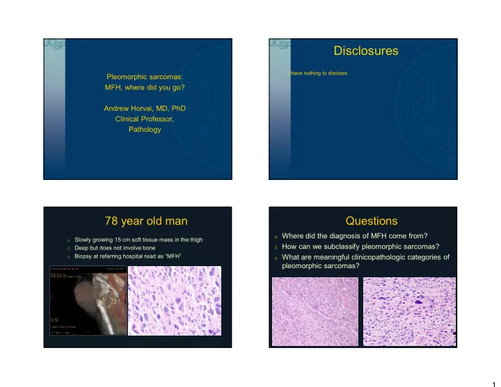
78 year old man

฀

Slowly growing 15 cm soft tissue mass in the thigh

฀

Deep but does not involve bone

฀

Biopsy at referring hospital read as “MFH”

Questions

฀ Where did the diagnosis of MFH come from? ฀ How can we subclassify pleomorphic sarcomas? ฀ What are meaningful clinicopathologic categories of

pleomorphic sarcomas?

slide-2
SLIDE 2

2

Where did MFH come from?

฀

1960s: Stout et al. multiple reports of “fibrous xanthoma” and “histiocytoma”

฀

Cultured cells from MFH (Ozello, Stout and Murray)

฀ Initially ameboid movement, phagocytosis  histiocytes ฀ Later, bipolar spindled cells  fibroblasts ฀ “Facultative fibroblasts”: histiocytes capable of fibroblast

differentiation

Kauffman SL, Stout AP. Cancer 1961 14:469-82. Ozzello et al Cancer 1963 16:331-44.

Where did MFH come from?

1972: Kempson and Kyriakos

Kempson RL, Kyriakos M. Cancer 1972 961-76.

MFH Histotype?

฀

Theories

฀ Facultative fibroblasts (facultative histiocytes?) ฀ Mixture of fibroblasts and histiocytes ฀ Stem cell with dual differentiation into fibroblasts and histiocytes

฀

Problems

฀ No histotype supported by ancillary diagnostic and in-vitro methods ฀ Some well-differentiated tumors (liposarcoma, leiomyosarcoma, etc.)

contain areas indistinguishable from pleomorphic, storiform or giant- cell MFH

฀ No unifying genetic changes (usually profound genomic instability)

“Histiocytes”
slide-3
SLIDE 3

3

Reappraisal of MFH

Fletcher CD. AJSP 1992 16:213-28. Fletcher CD et al J Clin Oncol 2001, 19:3045.

Myxofibrosarcoma Leiomyosarcoma Rhabdomyosarcoma Myogenic sarcoma NOS Pleomorphic liposarcoma WD liposarcoma Osteosarcoma Myofibroblastic sarcoma Fibrosarcomatous DFSP Evans tumor Other Undifferentiated sarcoma

Reappraisal of MFH (2001)

Fletcher CD et al J Clin Oncol 2001, 19:3045.

Undifferentiated sarcoma

Reappraisal of MFH (2001) Reappraisal of MFH (UCSF, 2017)

Dedifferentiated liposarcoma Pleomorphic rhabdomyosarcoma MPNST Adult fibrosarcoma Pleomorphic hyalinizing angiectatic tumor Myxoinflammatory fibroblasic sarcoma Undifferentiated sarcoma

slide-4
SLIDE 4

4

Reappraisal of MFH

  • Clinically meaningful classification, what is practical?
  • Not sarcoma
  • Melanoma
  • Carcinoma
  • Others: Germ cell, hematopoietic
  • Sarcoma (limited data)
  • Defined histotype
  • Dedifferentiated liposarcoma better than “MFH”
  • Myogenic sarcoma “worse” than non-myogenic
  • Uncertain histotype
  • Myxofibrosarcoma
  • Atypical fibroxanthoma / pleomorphic dermal sarcoma
  • Myxoinflammatory fibroblastic sarcoma
  • Undifferentiated pleomorphic sarcoma (UPS)

Pleomorphic tumors are not always sarcoma

Diagnosis Clin-path clues Immunostains Genetics Sarcomatoid carcinoma Cohesive cells, clinical history, superficial near epithelium Diffuse keratin P63+ INI1 intact Melanoma Nesting, in-situ component, pigment SOX10, S100, HMB45, MelanA BRAF V600E Germ cell tumor Young patient, para-aortic, mediastinum, history SALL4, OCT4, Keratin, CD30 Hematolymphoid In nodal anatomic sites, multifocal, round cells, dyshesive LCA, CD4, MPO, CD30 Variable

Sarcomatoid carcinoma

Sarcomatoid carcinoma

slide-5
SLIDE 5

5

Sarcomatoid carcinoma

Keratin EMA p63 INI1

Melanoma

T2 F/S T2 F/S Melanoma Melanoma

slide-6
SLIDE 6

6

Melanoma

S100 SOX10 MelanA

Pleomorphic sarcomas with defined histotype

Diagnosis Clin-path clues Stains Genetics Dedifferentiated liposarcoma Well differentiated component, retroperitoneum, scrotum MDM2, CDK4 Amp(12q13-15) Rhabdomyosarcoma Strap cells Myogenin, MyoD1, desmin Leiomyosarcoma Abundant dense cytoplasm Caldesmon, SMA, desmin (2 of 3) Myofibrosarcoma Loose, fasciitis-like SMA (tram-track) Pleomorphic liposarcoma Pleomorphic lipoblasts Oil red O Extraskeletal

  • steosarcoma

Produces osteoid or bone SATB2 (sensitive, not specific) Malignant peripheral nerve sheath tumor NF1, pre-existing benign nerve sheath tumor SOX10, S100 (sens. ~ 50%) H3K27Me3 loss

Pleomorphic sarcomas with defined histotype

McCormick et al Am J Surg Pathol 1994, 18:1213-23.

Dedifferentiated liposarcoma

slide-7
SLIDE 7

7

Dedifferentiated liposarcoma

Dedifferentiated Liposarcoma: 12q13-15 amplification

Horvai et al. 2009 Mod Pathol 22(S1): 14A

MDM2, CDK4

฀

Leiomyosarcoma

฀

Rhabdomyosarcoma

฀

Myofibroblastic sarcoma

฀

“Myogenic NOS”

Fletcher CD et al J Clin Oncol 2001, 19:3045.

Pleomorphic sarcomas with defined histotype

slide-8
SLIDE 8

8

Pleomorphic leiomyosarcoma Pleomorphic leiomyosarcoma

SMA Desmin Caldesmon Caldesmon

Pleomorphic rhabdomyosarcoma Pleomorphic rhabdomyosarcoma

slide-9
SLIDE 9

9

Pleomorphic rhabdomyosarcoma (myogenin) Myofibroblastic sarcoma Myofibroblastic sarcoma

Myofibroblastic sarcoma

SMA Desmin Caldesmon Caldesmon

slide-10
SLIDE 10

10

Pleomorphic sarcomas with defined histotype - clinical

Diagnosis Recurrence Metastasis Mortality Dedifferentiated liposarcoma 51% 15% 26% @ 5 yrs. Myogenic sarcomas ?% 56% 39% @ 20 yrs. Undifferentiated pleomorphic sarcoma 13-42% 31-35% 37% @ 5 yrs.

McCormick et al Am J Surg Pathol 1994, 18:1213-23. Fletcher CD et al J Clin Oncol 2001, 19:3045.

Pleomorphic sarcomas with uncertain histotype

Diagnosis Clin-path clues Stains Genetics Myxofibrosarcoma Superficial, predominantly myxoid, branching vessels Nonspecific, most negative Variable Atypical fibroxanthoma / Pleomorphic dermal sarcoma Dermal, subcutaneous, sun damage Nonspecific: Vimentin, CD10; negative for keratin and melanocytic NOTCH1/2 FAT1 Pleomorphic hyalinizing angiectatic tumor Foot, middle age, damaged vessels, 0-1/10 hpf S100 negative TGFBR3-MGEA5 Myxoinflammatory fibroblastic sarcoma Acral, foot, pseudolipoblasts, inflammation, <1 mf / 50 hpf Nonspecific BRAF (in pure MIFS)

Myxofibrosarcoma (myxoid “MFH”) Myxofibrosarcoma

slide-11
SLIDE 11

11

Atypical fibroxanthoma, undifferentiated dermal sarcoma Atypical fibroxanthoma, pleomorphic dermal sarcoma

McCalmont TH. J Cutan Pathol 2011; 38:853. McCalmont TH. J Cutan Pathol. 2012 39:8.

Pleomorphic hyalinizing angiectatic tumor (PHAT) Myxoinflammatory fibroblastic sarcoma (MIFS)

slide-12
SLIDE 12

12

Myxoinflammatory fibroblastic sarcoma (MIFS) Pleomorphic sarcomas with uncertain histotype - clinical

Odei B et al. Am J Clin Oncol 2017 epub. Koch M et al. Anticancer Res 2015 35:5717. Miller K et al. Am J Surg Pathol 2012 36:1317. Folpe AL, Weiss SW. Am J Surg Pathol 2004 11:14-17. Laskin WB et al. Am J Surg Pathol 2014 38:1. Roland CL et al. Ann Surg Oncol 2016 23:2220.

Diagnosis Recurrence Metastasis Mortality Myxofibrosarcoma (grade 1-3) 31% 15% 23% @ 3 yrs. Atypical fibroxanthoma 8% 0-3% 0-2% @ 20 yrs. Pleomorphic dermal sarcoma 28% 5-10% 0-2% @ 2 yrs. Pleomorphic hyalinizing angiectatic tumor 33% 0% 0% @ 4 yrs. Myxoinflammatory fibroblastic sarcoma 51% 2% 0% @ 5 yrs. Undifferentiated pleomorphic sarcoma 13-42% 31-35% 37% @ 5 yrs.

How much further should we subdivide UPS?

Undifferenti- ated sarcoma

฀

Lymphoma: DLBCL, anaplastic

฀

Myeloid sarcoma

฀

Follicular “dendritic cell”

฀

Interdigitating “dendritic cell”

฀

True histiocytic sarcoma

Cannatella J and Horvai A. Manuscript in prep

How much further should we subdivide UPS?

Undifferent iated sarcoma

฀

Lymphoma: DLBCL, anaplastic

฀

Myeloid sarcoma

฀

Follicular “dendritic cell”

฀

Interdigitating “dendritic cell”

฀

True histiocytic sarcoma

Cannatella J and Horvai A. Manuscript in prep

slide-13
SLIDE 13

13

What do I do?

฀

Biopsy:

฀ Desmin, SOX10, Keratin ฀ History of NF1: S100, H3K27Me3 ฀ Looks myoid: SMA, caldesmon ฀ If retroperitoneal, scrotal or imaging to suggest fat: MDM2, CDK4

฀

Excision:

฀ H&E: lipoblasts, cartilage, bone, benign neurofibroma, mitotic activity ฀ Same stains as above ฀ Make sure mitoses correlate with pleomorphism esp. in feet

Take-home messages

  • 1. Avoid the diagnosis of Malignant Fibrous Histiocytoma

(MFH).

  • 2. Unclassified Pleomorphic Sarcoma (UPS) is a diagnosis of

exclusion.

  • 3. Prior to diagnosing UPS, exclude non-sarcomas, myogenic

sarcoma, dedifferentiated liposarcoma.

  • 4. Consider AFX/PDS based on dermal location