Esophageal Motor Abnormalities Brooks D. Cash, MD, FACP, AGAF, FACG, - - PDF document

esophageal motor abnormalities
SMART_READER_LITE
LIVE PREVIEW

Esophageal Motor Abnormalities Brooks D. Cash, MD, FACP, AGAF, FACG, - - PDF document

6/2/2015 Esophageal Motor Abnormalities Brooks D. Cash, MD, FACP, AGAF, FACG, FASGE Professor of Medicine Gastroenterology Division University of South Alabama Mobile, AL High Resolution Manometry Pivotal Advance Late Ray Clouse, MD


slide-1
SLIDE 1

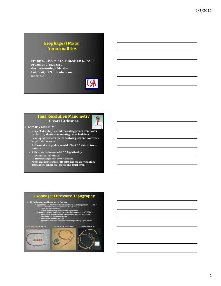
6/2/2015 1 Esophageal Motor Abnormalities

Brooks D. Cash, MD, FACP, AGAF, FACG, FASGE Professor of Medicine Gastroenterology Division University of South Alabama Mobile, AL

High Resolution Manometry Pivotal Advance

  • Late Ray Clouse, MD

– Suspected widely spaced recording points from water perfused systems were missing important data – Developed spatiotemporal contour plots and converted amplitudes to colors – Software developed to provide “best fit” data between sensors – Solid state catheters with 36 high‐fidelity circumferential sensors

  • Entire esophagus could now be visualized

– Additional refinements (3D HRM, impedance, video) and applications (anorectal, gastric and small bowel)

Esophageal Pressure Topography

  • High‐Resolution Manometry Catheter
  • Spans from the pharynx to the stomach with sensor separation of no more

than a centimeter within and around the sphincters.

– Greater than 32 pressure sensors – Temporal frequency response matched to the zone of the esophagus

  • Compared to water perfusion, the immediate advantages of HRM are:

1) simplified procedural set up with improved sphincter localization

2) elimination of movement artifact

3) simplified data interpretation

4) ability to perform more sophisticated analysis of esophageal function.

Each sensor has 12 pressure sensitive segments

Sierra Scientific Instruments Medical Measurement Systems Sandhill Scientific Inc.

slide-2
SLIDE 2

6/2/2015 2

Manometric port

40 mmHg 2.5 4.5 5.6 7.1 8.5 13.0 8.8 11.0

Functional Imaging of Esophageal Peristalsis

HIGH‐RESOLUTION MANOMETRY

Clouse Plot

Manometric port

NU IRB

2.5 4.5 5.6 7.1 8.5 13.0 8.8 11.0

True Functional Imaging of Esophageal Peristalsis

ESOPHAGEAL PRESSURE TOPOGRAPHY

‐10 10 30 70 90 ≥110 50

Pressure mmHg

Clouse Plot

slide-3
SLIDE 3

6/2/2015 3

Pressure Topography of Esophageal Motility: What does it add?

  • More akin to an imaging modality

– Defines important anatomical landmarks and abnormalities – Refines measurement of important motor events

  • EGJ relaxation
  • Peristaltic timing velocity
  • Contractile activity/force/amplitude

– Defines intra‐luminal pressurization patterns – Permits pattern recognition

3 Main Steps in Diagnostic Approach to a High Resolution Manometry Test

  • 1. Assess EGJ anatomy and function
  • 2. Assess esophageal body function
  • 3. Review pressurization patterns

These 3 steps will permit diagnosis of most esophageal motor abnormalities * Some changes in prioritization with recent Chicago Classification update (v3.0)

Anatomy of a High Resolution Esophageal Manometry Test

slide-4
SLIDE 4

6/2/2015 4

STEP 1| Assess the EGJ Anatomy and Function

  • Determine if hiatus hernia is present
  • Confirm that the catheter has crossed the EGJ and diaphragm

Integrated relaxation pressure: The IRP will determine whether

  • utflow obstruction at the EGJ is evident. Disorders are separated

at this point, determined by those with or without outflow

  • bstruction at the EGJ.

DEEP BREATH

3

Integrated Relaxation Pressure (IRP): Mean EGJ pressure measured with a sleeve for 4 contiguous or non‐contiguous seconds of relaxation in the 10‐second window following deglutitive UES relaxation.

  • The upper limit of normal using ManoScan is 15 mmHg.

IRP INTEGRATED RELAXATION PRESSURE IRP INTEGRATED RELAXATION PRESSURE

Assess the EGJ Anatomy and Function

slide-5
SLIDE 5

6/2/2015 5

STEP 2 | Assess Esophageal Body Function

  • Peristaltic integrity: either intact, weak or failed
  • Contractile deceleration point (CDP): anatomic separation

point (between tubular esophagus and phrenic ampulla)

  • Distal Latency (DL): timing of esophageal peristalsis
  • will define the swallow as premature or normal latency
  • Distal contractile index (DCI): vigor of the distal

esophageal contraction

  • Contractile front velocity (CFV): speed of esophageal

contractions

  • previously used to define rapid contraction
  • no longer considered meaningful

Peristaltic Breaks: Gaps in the 20 mmHg isobaric contour of the

peristaltic contraction between the UES and EGJ, measured in axial length.

LARGE BREAK FAILED SWALLOW

Contractile Deceleration Point (CDP): The inflection point along the 30 mmHg

isobaric contour where propagation velocity slows, demarcating the tubular esophagus from the phrenic ampulla.

Distal Latency (DL): Interval between UES (1) relaxation and the CDP (2), expressed in

  • seconds. Normal DL is >4.5 sec.

CDP CONTRACTION DECELERATION POINT (2) UES RELAXATION (1) CDP CONTRACTION DECELERATION POINT (2) DL DISTAL LATENCY CDP CONTRACTION DECELERATION POINT (2)

DL: 7.8 sec

slide-6
SLIDE 6

6/2/2015 6

STEP 3 | Pressurization Patterns

Each swallow should be evaluated using the IBC tool to document an isobaric pressurization above 30 mmHg.

Achalasia: HRM led to the identification of three discernible achalasia types. Each subgroup represents a distinct clinical entity, each with significantly different biomechanics and treatment outcomes. Type I patients do significantly better with Heller myotomy than with pneumatic dilatation, and Type III patients exhibit the worst prognostic outcome.

Achalasia TYPE I

There is no significant pressurization within the body

  • f the

esophagus. Therefore, this would be classified as failed peristalsis with abnormal IRP . In the absence of esophageal body contractility, the IRP threshold of >10 mmHg is used to distinguish Type I Achalasia from absent peristalsis.5

Failed peristalsis with abnormal IRP - no esophageal function

Major Disorders of Esophageal Peristalsis

  • Achalasia
  • Hypertensive LES/EGJ Outflow obstruction
  • (Nutcracker esophagus)
  • Jackhammer esophagus
  • Distal esophageal spasm (DES)
  • Absent peristalsis

Pressure Topography of Esophageal Motility

The Chicago Classification

Neurogastroenterology and Motility, 2015;27;160‐74.

slide-7
SLIDE 7

6/2/2015 7 Chicago Classification 3.0 Changes

  • Use median rather than mean cutoff value for IRP
  • Use lower IRP cutoff for type I achalasia (platform specific)
  • Panesophageal pressurization with ≥ 20% swallows with

100% failed contractions is type II achalasia irrespective of IRP

  • Emphasize heterogeneity of conditions potentially causing

EGJ outflow obstruction

  • Modify hypercontractile esophagus to ≥20% swallows with

DCI >8000 mmHg x s x cm

  • Substitute ‘absent contractility’ for ‘aperistalsis’ or ‘absent

peristalsis’ to differentiate from other scenarios where peristalsis is absent (e.g., achalasia)

  • Rename ‘minor disorders of peristalsis’
  • Eliminate small breaks (2–5 cm) in the 20‐mmHg isobaric

contour as a criterion of abnormality

  • Eliminate rapid CFV (>9 cm/s) as a criterion of abnormality
  • Eliminate the designation of ‘hypertensive peristalsis’ (DCI

5000–8000 mmHg x s x cm) (no more Nutcracker)

  • Adopt the ‘ineffective esophageal motility’ (IEM)

designation from conventional manometry

  • Eliminate ‘frequent failed peristalsis’ as a distinct

diagnostic entity

  • Incorporate new data from studies of multiple repetitive

swallows into the criteria for IEM

Chicago Classification 3.0 Changes

EGJ Outflow Obstruction

  • Incompletely expressed

achalasia

  • Mechanical obstruction

IRP ≥ upper limit of normal AND some instances of intact or weak peristalsis Y es

A c halasia

  • Subtype I: No contractility
  • Subtype II: ≥ 20% PEP
  • Subtype III: ≥ 20% spasm

(DL<4.5s)

IRP ≥ ULN AND 100% failed peristalsis or spasm Y es No

Neurogastroenterology and Motility, 2015;27;160‐74.

Disorders with EGJ Outflow Obstruction

The Chicago Classification

slide-8
SLIDE 8

6/2/2015 8

Achalasia

  • Dysphagia, wt loss,

regurgitation, halitosis, GERD sxs

  • Immune‐mediated disease

targeting esophageal myenteric plexus (neurons and ganglia)

– Antineuronal Abs, inflammatory cells, cytokines, immunoglobulins, complement – Achalasia subtypes may represent differential degree

  • f immune

activation/selectivity (cell vs humoral) – HSV‐1 implicated as trigger

Kahrilas PJ, et al. Gastroenterology 2013;145:954‐66.

100 50 mmHg 150 30

Type I Type II Type III

IRP= 22.3 mmHg IRP= 24.2 mmHg IRP= 29.8 mmHg air liquid air EGJ EGJ EGJ 5 seconds 5 seconds 5 seconds

High‐Resolution Manometry: Achalasia subtypes

contraction diverticulum

Clinical Evolution of Achalasia

Assessing clinically relevant phenotypes

Early Type II or III Late Type I Chronic Type II/III‐‐I

NU IRB

slide-9
SLIDE 9

6/2/2015 9

Achalasia Mimics

  • Malignancy (Pseudoachalasia)
  • Chaga’s disease
  • Amyloidosis
  • Postvagotomy
  • Neurofibromatosis
  • Sarcoidosis
  • MEN IIb

Hypertensive LES

  • Presentation: Chest pain/dysphagia/globus

– May be an achalasia variant

  • Dx: LES pressure > 35 mmHg AND failure to relax

below IRP of 15 mmHg

– Normal peristalsis

  • More important than pressures: failure of full

relaxation at LES

– Incomplete bolus transfer

  • Can overlap with other spastic esophageal

conditions

– May need additional provocation (bread swallow, multiple rapid swallows, solid swallows) – EUS recommended prior to therapy to exclude infiltrative

  • r compressive disease (eg malignancy)

A:EGJOO:achalasia phenotype B:EGJOO: Mechanical

100 50 mmHg 150 30 IRP= 22.3 mmHg IRP= 27.2 mmHg Barium tablet localized 12 mm restriction EGJ Large diverticulum 4 cm above EGJ Locus of diverticulum above EGJ Compartmentalized pressurization Normal peristalsis

EGJ Outflow Obstruction

slide-10
SLIDE 10

6/2/2015 10

DES

  • ≥ 20% of swallows with reduced DL(<4.5s)

Jackhammer esophagus

  • DCI > 8,000 mmHg‐s‐ cm and normal DL

Absent contractility IRP is normal AND short DL or high DCI or 100% failed peristalsis

Major Disorders of Peristalsis

The Chicago Classification

Y es

Neurogastroenterology and Motility, 2015;27;160‐74.

Hypercontractile Esophagus The diagnostic criteria for

hypercontractile peristalsis have been redefined as at least one propagated swallow‐induced contraction with a DCI of >8,000 mmHg‐s‐cm, as that value is extremely rare in asymptomatic subjects. The repetitive contractions sometimes seen in this presentation have led to the reference of “Jackhammer” esophagus.

Distal Esophageal Spasm Distal Latency (DL) has been proposed

as an improved measure to represent simultaneous contractions. The DL is currently used to define DES, although further evaluation of clinical

  • utcomes is needed to support this metric.1

Borderline Esophageal Motor Function This category

represents a range of abnormalities including borderline‐normal to those conditions similar to ineffective esophageal motility (IEM) or absent contractility.

Non‐achalasia motor abnormalities Nutcracker Esophagus

  • Prolonged hypertensive contractions with normal

swallow waveform propagation

  • Etiology: possible hypercholinergic state resulting in

incoordination of longitudinal and smooth muscle

  • Presentation: episodic chest pain (may be mistaken

with GERD or cardiac CP)

– Dysphagia (solids or liquids) – Exacerbation with hot or cold – ENT: High dysphagia (globus)

  • May exist with other abnormalities (LES HTN)
  • Dx: DCI > 5000 (cont ampl x duration x length)
slide-11
SLIDE 11

6/2/2015 11

Hypercontractile or Jackhammer Esophagus (aka Spastic Nutcracker)

  • Offshoot of Nutcracker

– 4% manometry referrals; r/o mechanical obst

  • Presentation: Chest pain/dysphagia
  • Dx:

– At least one DCI > 8000 – Repeated high amplitude contractions – Normal DL (≥ 4.5 sec)

Distal Esophageal Spasm (DES)

  • Unknown etiology; likely related to defects

in inhibitory neural pathways of esophagus

– Rarest manometric diagnosis (3%) – Classic corkscrew esophagus very rare

  • Presentation: Chest pain/dysphagia

– Symptom correlation poor

  • Dx:

– Normal median IRP (LES relaxation), ≥ 20% premature contractions with DCI > 450 mmHg x s x cm – Some normal peristalsis may be present

21 19 17 15 13 11 9 7 5 3 1 0s 10s 5s A:Latency described with conventional manometry B: Latency measured with EPT Normal swallow Axial position (cm) CDP DL

Time (s)

CFV he ) t mc

  • ng us

( al h

  • phag

t Leng es 100 50 30 mmHg 150 10 Length along the esophagus (cm) 30 35 5 15 1 20 25 C: DES: Rapid Premature Contraction

DL = 4.4 s CFV = 6 cm/s

2 s

Time (s) 5 10 15 20 25 30 35 Bolus Escape 3.0 cm 30 cm/s

DL = 3.0 s CFV = 45 cm/s

2 s

Time (s) DL = 7.0 s CFV = 15 cm/s 5.5 cm D: DES: Premature Contraction E: Rapid Contraction with Normal latency

2 s

Time (s) 100 50 30 mmHg 150

slide-12
SLIDE 12

6/2/2015 12

UES EGJ

10 s 5 10 100 15 20 25 30 35 Length along the esophagus (cm) EGJ relaxation A) Normal: No breaks/ NL DCI 50 30 150 mmHg DCI= 20,452 mmHg-s-cm DCI= 2,761 mmHg-s-cm B) Jackhammer: No breaks/ Abnormal DCI DCI= 34 mmHg-s-cm 5 10 15 20 25 30 35 Length along the esophagus (cm) 10 s 10 s 15 s 10 s 10 s DCI= 7 mmHg-s-cm DCI= 1121 mmHg-s-cm C) Absent Peristalsis D) Absent/Failed Peristalsis E) Weak Peristalsis- IEM F) Weak Peristalsis- TZ Defect DCI= 325 mmHg-s-cm

Treatment of Achalasia

Response Rates of Achalasia Treatments

Patients categorized by pressure topography subtype

Pandolfino JE, et al. Gastroenterology 2008 Salvador R, et al. J Gastrointest Surg 2010 Pratap N, et al. J Neurogastroenterol Motil 2011 Rohof W, Gut 2011; 60 (Suppl 3)

slide-13
SLIDE 13

6/2/2015 13

Treatment of Achalasia: 2015

  • POEM (Per‐oral endoscopic myotomy)

– Short‐term success 82‐100% – LES pressures reliably lowered – Lack of long‐term data/comparisons to laparascopic Heller myotomy (current data suggests equivalence)

Treatment of other Esophageal Hypertensive Conditions

  • Nutcracker/Jackhammer

– Trial of nitrates (SL or oral) + PPI – Calcium channel blockers (diltiazem, sildenafil) PRN – Tricyclic antidepressants

  • Hypertensive LES/EGJ Outflow obstruction

– Balloon dilation or botox injection – SAA – POEM

  • DES

– PPI + Botox injection – Surgical myotomy (when all else fails)

POEM for Spastic Disorders

  • 73 patients

– 9 DES; 10 Jackhammer, 54 type III (spastic) achalasia – Dysphagia, regurg, chest pain – 11 medcens

  • 100% completion

– 118 mins (mean) – 19 cm tunnel (mean) – 16 cm myotomy (mean) – 3.4 hospital days (mean)

  • 8 AEs (11%) none severe

Khashab M, et al. Gastrointest Endosc 2015;81:1170‐7

slide-14
SLIDE 14

6/2/2015 14 POEM for Spastic Disorders

Khashab M, et al. Gastrointest Endosc 2015;81:1170‐7

  • There is no clear discriminator of symptomatic

hypercontractility

  • Propagation can appear normal
  • Therapy focused on reducing peristaltic

amplitude in altering symptoms is extremely limited

  • Smooth muscle relaxants
  • BOTOX
  • Treat visceral sensitivity

Hypertensive Contractility Caveats

Ineffective motility

  • ≥ 50% ineffective swallows

Fragmented Peristalsis

  • ≥ 50% fragmented swallows with large (>5 cm)

breaks in the 20-mmHg IBC and not effective

IRP is normal AND ≥ 50% ineffective swallows

Minor Disorders of Peristalsis Non‐hypertensive Esophageal Motility Disorders

The Chicago classification

Y es

Minor Disorders of Peristalsis: Impaired clearance

Neurogastroenterology and Motility, 2015;27;160‐74.

slide-15
SLIDE 15

6/2/2015 15

Minor Disorders of Peristalsis

Neurogastroenterology and Motility, 2015;27;160‐74.

Symptoms of dysphagia ± chest pain and bland regurgitation Upper Endoscopy

Obstructive process: ring, stricture, etc. Normal Esophageal dilatation EGJ resistance Retained food Diverticulum

High Resolution Manometry

*esophagram may be helpful when manometry is technically difficult to perform

Utilizing HRM/EPT in the Management of Esophageal Symptoms

EGJ Outflow Obstruction Absent Contractility Achalasia I

  • Severe dilatation

associated with poor treatment response

Achalasia II

  • Best treatment

response

Achalasia III

  • Worst treatment

response

DES

  • Extremely rare
  • EGD ± EUS/CT to rule
  • ut obstructive

process

  • If clinical scenario c/w

achalasia, a timed barium esophagram should be performed

  • Consider myotomy as

initial therapy

  • Esophagram can be

normal without barium retention or esophageal dilatation

  • May benefit from

treatment directed at spasm

  • Difficult to treat
  • Many cases

misdiagnosed Type III achalasia

  • Potentially achalasia

phenotype with preserved peristalsis

  • Potentially advanced

GERD or scleroderma

  • Potentially achalasia

phenotype with hypotensive LES Frequently misdiagnosed with conventional manometry Often diagnosed as DES

  • n esophagram

Key Take Home Points

– Esophageal symptoms can have a number of

  • verlapping etiologies

– Interaction between organic and functional influence should not be ignored – Most disorders can be managed by a careful systematic evaluation

  • Exclude most dangerous causes first and then focus on the

most likely cause

– Diagnose and treat in parallel – Embrace the evolving technology available to you

Approach to Patients with Esophageal Symptoms