How to Select Patients for Treatment Thermal tumescent of Chronic - - PowerPoint PPT Presentation

how to select patients for treatment
SMART_READER_LITE
LIVE PREVIEW

How to Select Patients for Treatment Thermal tumescent of Chronic - - PowerPoint PPT Presentation

Chronic Venous Disease Treatment Options Rigid or short stretch compression(Unnas boot or Profore) Endovenous ablation of the saphenous vein How to Select Patients for Treatment Thermal tumescent of Chronic Venous Insufficiency


slide-1
SLIDE 1

1

How to Select Patients for Treatment

  • f Chronic Venous Insufficiency

Peter F. Lawrence, MD Professor and Chief Division of Vascular Surgery University of California Los Angeles

Chronic Venous Disease Treatment Options

Rigid or short stretch compression(Unna’s boot or Profore) Endovenous ablation of the saphenous vein – Thermal tumescent – Nonthermal non-tumescent Microphlebectomy of tributary veins Sclerotherapy of tributary veins Perforator ablation (SEPS,RFA/Laser, Sclero Iliac venous stent Cross-femoral venous bypass Femoral-popliteal venous bypass

Lower Extremity Venous Anatomy

Superficial Vein System Perforator Vein System

slide-2
SLIDE 2

2

Pathophysiology of CVI

Vein valves

– Bicuspid valves – Blood propelled by calf muscle pump opens the valve in one direction – Blood moving with gravity closes the normal valve – Incompetence of valve at saphenofemoral junction is the source of most varicose veins

Proximal Iliac Vein Stenosis/Obstruction Proximal Iliac Vein Stenosis/Obstruction

Prospective study of 78 CEAP 5/6 patients to assess incidence

  • f proximal venous disease

– Imaging modality: Duplex

ultrasound plus combined with either CTV or MRV

– M:F = 50%; mean age =60 – Ulcers equally distributed

between R and L

– 50% had hx of DVT

37% had iliocaval stenosis of >50%; 23% had stenosis/occlusion>80% Independent risk factors=

– Women – Hx of DVT – Deep venous reflux

JVS 2011;53:1303-1308

Chronic Venous Insufficiency Classification

Clinical

– (1) Telangiectasias – (2) Varicose veins – (3) Edema – (4) Skin changes – (5) Ulcer-healed – (6) Ulcer-active

Etiologic

– Primary – Secondary – Congenital

Anatomic

– Deep – Superficial – Perforator

Pathophysiologic

– Reflux – Obstruction – Reflux + obstruction

JVS 2004;40(6):1248-1252

slide-3
SLIDE 3

3

Classification System

CEAP is the best classification system The most common chronic disease in the US

– 40% of Americans have venous disease – 15-25% of the adult population have saphenous or branch incompetence – 40% of adults have telangiectasias (spider veins)

One of the most common causes of wounds

– 6% have a venous ulcer during their lifetime

Ann Epidemiol 2005;15(3):175-184 Circulation 2005;111:2398-2409

Assessing Venous System Non-invasive vascular lab

Duplex scan in reversed Trendelenberg position to assess for:

– Great/Small saphenous incompetence Large vein > 3.5 mm > 0.5 sec reflux – Perforator vein incompetence Large vein > 3.5 mm >0.5 sec reflux Pulsatility – Tributary incompetence – Presence of deep vein

  • bstruction/incompetence

JVS 2014;60:Supplement S

Support hose, Circaid, Short Stretch Compression Bandage and Unna’s Boot Heat Inducing Devices That Require Tumescence

Superficial Veins Laser and Radiofrequency Perforator Veins RFA approved; Sclero and laser also used

slide-4
SLIDE 4

4

Radiofrequency and Laser Ablation Mechanism of Action

2-3 cm

Perforator Ablation (Difficult!)

RFS catheter placed at a 45 degree angle- transverse and longitudinal transducer Confirm stylet in vein at fascia Inject local anesthetic to eliminate pain Trendelenberg position Vein treated with radiofrequency energy for 1 minute in each quadrant; repeated above the fascia if possible Confirmation of ablation post procedure difficult due to compression from local anesthetic

Single Surgeon Learning Curve

JVS 2010;54(3):737-742

Learning curve of a Single Vascular Surgeon 0% 10% 20% 30% 40% 50% 60% 70% 80% 2007 2008 2009 2010 success rate

Perforator Closure Success Rate

0% 10% 20% 30% 40% 50% 60% 70% 80% 2007 2008 2009 2010

success rate

Stab Phlebectomy Procedure

Incisions (Nokor Needle)

– Adjacent to the vein to avoid cutting it – Length no greater than the crochet hook width (1 mm) – Penetrate dermis only

Loop of vein pulled through incision Mosquitoes used to divide vein Circular motion used to remove the vein- limits the size of the incision More incisions better than larger incisions Vein path compressed with finger to reduce hematoma Transillumination can be used for complete vein removal

slide-5
SLIDE 5

5 CEAP 1 Patients Telangiectasias or reticular veins

Elective – Cosmetic Treated with injection (sclerotherapy) or laser

JVS 2014;60:Supplement S

CEAP 2 Patients Varicose veins

Goals for Treatment

– Elimination of reflux and any incompetence vein(s) – Reduce pain – Limited anesthesia – Short recovery – Excellent cosmetic result – Low complication rate – Low recurrence rate

SVS-AVF guidelines: compression of 20-30 mmHg recommended, knee or thigh high (Grade 1, Level B)

JVS 2014;60:Supplement S

CEAP 2 Patients Varicose veins – Treatment Options

Sclerotherapy Microphlebectomy

Endovenous laser ablation of GSV/SSV Radiofrequency ablation of GSV/SSV Nonthermal,nontumescent Mechanico-chemical Rotational wire/Sclero/Glue

CEAP 2 Patients Varicose veins

Ann Surg. 2011;254(6)876:881

3-month technical success

– 96% EVLT – 97% RFA – 95% NT,NT Postop pain was considerably lower for RFA Changes in quality of life between pre and post-operative were not significantly different

slide-6
SLIDE 6

6 CEAP 3 Patients Edema

Patients with edema, pain, and/or varicose veins Compression therapy* Resolution of symptoms Duplex ultrasound of superficial veins No resolution of symptoms Treatment of incompetent superficial veins *For post-thrombotic patients the SVS-AVF recommends compression of 30-40, knee or thigh high (Grade 1, Level B)

JVS 2014;60:Supplement S

Impact of Superficial Vein Ablation for CEAP 2-4 Patients

Presenting Symptoms Number of patients Lifestyle limiting pain n=696 (94.8%) Limb swelling n=485 (66%) Lipodermatosclerosis n=39 (5.3%) Bleeding varicose veins n=10 (1.4%)

Impact of Superficial Vein Ablation for CEAP 2-4 Patients

Clinical Severity Number of Limbs Relief of Symptoms (Mean=9 months) Symptomatic varicose veins (C2) 217 92% Swelling (C3) 346 91% Hyperpigmentation and/or Lipodermatosclerosis (C4) 230 81%

98.6% closure success rate at 24-72 hr Mean follow-up = 9 mo. Late GSV recanalization = 1.8% Late SSV recanalization = 0%

Mid-Advanced CVI CEAP 4-6 Patients

When to Intervene Patient presents with a leg that is pigmented and fibrotic in the “gaiter zone” of the ankle Patient presents with non- healing ulcer that persists in spite of optimal compression Patient presents with a healed ulcer at the location of an incompetent perforator vein

slide-7
SLIDE 7

7

What To Do With Patients with Progressive Non Healing Venous Ulcers?

Correct the ambulatory superficial venous hypertension by:

– Great saphenous ablation- agreed – Small saphenous ablation-agreed – Tributary eradication-agreed – Perforator ablation - ???? Linton procedure Too many wound infections SEPS Difficult to get ankle perforators Laser/RFA ablation Sclerotherapy Deep system valvuloplasty/bypass – not routinely used

  • r

Compression for life– and 25% with ulcers have weekly wound care!

SVS-AFV Practice Guidelines Operative/Endovascular - Algorithm

Venous Ulcer

Diagnostics

+ reflux + obstruction

  • + proximal

Rx Endo Failed/ not option Reevaluate + proximal + infrainguinal

Bypass unilateral +/+ +/- Bypass/ endophlebectomy

  • /+

Not option/ Reevaluate

Bypass bilateral

+ perforator + superficial Rx Endo (open) ***

*** clarification: +/+ ulcer treat both, no ulcer treat only superficial

Failed/ not option Reevaluate

+ Superficial/perforator Deep reflux Valvular reconstruction

similar risk/benefit: Less risk

similar risk/benefit: Moderate risk Highest risk

Simplified Patient Algorithm CEAP 4- 6 Patients CEAP 5 Patients Healed venous ulcer

Guiding Information

– Did the patient have difficulty healing the ulcer with compression bandages? – Is the patient compliant in wearing appropriate support stockings? – Does the patient have any incompetent superficial or perforator veins? – Does the patients lifestyle increase their risk of recurrence?

slide-8
SLIDE 8

8

Is Compression Enough to Prevent Ulcer Recurrence?

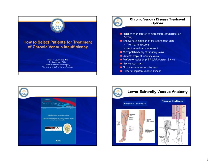
Patients with compression and ablation of incompetent superficial and perforator veins had lower ulcer recurrence rate to historic controls of compression alone Patients whose ulcer recurrences often have new incompetent perforator veins in which ablation can lead to ulcer healing

STUDY PATIENTS (n =) THERAPY 1 YR RR 2 YR RR 3 YR RR

Gohel et al1 500 Compression 28% N/A 56% Erickson et al2 71 Compression 35% 42% 49% Harlander-Locke et al3 20 Compression + Ablation 5% 10% 10%

(1) JVS, 2007 (2) JVS, 1995 (3) JVS, 2012

Compression therapy remains the mainstay treatment for a majority of patients

– Rigid or elastic wrap (3 months)

Eliminate superficial reflux

– Saphenous, small saphenous, accessory saphenous – Eliminate tributary reflux

Eliminate perforator reflux

– Region of ulcer and above the ulcer

Compression to accelerate wound healing Adjuncts

– Skin grafts – Skin substitutes – Growth factors/stem cells

CEAP 6 Patient Active venous ulcer Compression Therapy Does Not Cure All Venous Ulcers Alone

2 yrs = 17%, 3 yrs = 22% The difference between compliant and non-compliant groups was significant, but 22%

  • f the compliant patients had

progression/recurrence

Moneta et al, 1996

Track impact of current treatment

– Measurement of size of ulcer- if no improvement move to next treatment

Radiofrequency Ablation (great and small saphenous veins)

– Compression for 3 months – Measurement of size of ulcer

Ablation of perforators immediately adjacent to the ulcer, if ulcer is stable or enlarging Ablation of other adjacent ulcers if continued ulcer growth

CEAP 6 Patient Active venous ulcer

slide-9
SLIDE 9

9

Method of Ulcer Evaluation

0.00 2.00 4.00 6.00 8.00 10.00 12.00 14.00 16.00 0.00 0.10 0.20 0.30 0.40 0.50 0.60 Ulcer Size (cm^2) Time (Years) Ulcer growth = +2.66 cm2/mo Ulcer closure =

  • 6.08 cm2/mo

Green: Treatment with compression therapy alone Orange: Ongoing compression after vein ablation

Vein Ablation Procedure

Where Does Superficial and Perforator Ablation Fit in the Treatment of CVI?

O’Donnell 2008

– Few randomized trials(4) – ESCHAR trial (2007) showed no improved ulcer healing, but reduced ulcer recurrence when superficial veins treated- no perforators Rx’ed

GSV stripping or ablation results in reduced ulcer recurrence- Level 1-A evidence

JVS 2008;48:1044-1052

Impact of Perforator Ablation Randomized Control Trials

Current State of the Treatment of Perforating Veins. JVS 2015. In-Press

Ablation of incompetent perforator veins resulted in a mean of +4% ulcer healing Ablation of incompetent perforator veins resulted in a mean of -13% ulcer recurrence

Choosing the Right Method for Perforator Ablation

Ultrasound guided foam sclerotherapy (UGFS) = 65% Radiofrequency = 83% Laser = 75% SEPS = 87%- no longer used

Mean Success Rate

*Studies in last 5 years Current State of the Treatment of Perforating Veins. JVS 2015. In-Press

slide-10
SLIDE 10

10

Single Institution Studies on Perforator Ablation for Ulcer Healing

Mean ulcer healing = 81% Mean ulcer recurrence = 10%

Current State of the Treatment of Perforating Veins. JVS 2015. In-Press

Why Have Some Studies Shown No Benefit From Perforator Ablation?

Diagnosis

– Inaccurate Identification of Incompetent Perforators

Treatment sequence

– Inappropriate Sequencing of Treatment of CVI Superficial veins first, then tributaries, then perforators

Technique

– Poor Technique in Performing Perforator Ablation – Ablation of Wrong Perforators

Measurement of progress

– Inadequate Measurement of Ulcer Area and Volume

Incomplete Compression and Inadequate Wound Care

Proximal Venous Outflow Obstruction

Raju’s Criteria for Proximal Venous Stenting

One or more of the following clinical symptoms:

– Diffuse venous (orthostatic) limb pain that is graded >3/10 in visual analogue scale or interfering with sleep or work or requiring regular analgesic/narcotic use. – Venous leg swelling that is grade 3 (more than ankle edema involving the calf. Very rarely lesser degrees of swelling may be considered if it is combined with significant pain. – Venous stasis dermatitis/hyperpigmentation/lipodermatosclerosis – Venous stasis ulceration either active or healed if recurrent

Comment: If saphenous reflux is present, a saphenous ablation is generally undertaken before considering venous stenting

slide-11
SLIDE 11

11

Impact of Venous Outflow Obstruction

Post-stenting changes

– Decrease in ambulatory venous pressure – Reduction in swelling* – Reduction in pain* – Half of venous ulcers healed

*Significant (P<.0001) JVS 2003;38:879-885

Conclusions from Literature CEAP 5 – 6 Patients

Venous ulcers can be healed with an aggressive approach to incompetent superficial axial veins and perforating veins Healed venous ulcers (CEAP 5) can be maintained with a combination of compression and ablation of incompetent axial and perforator veins The status of the ulcer is the key to determining if there is persistent “ambulatory venous hypertension” If an ulcer is not healing with optimal compression (CEAP 6) or heals and has progressive lipodermatosclerosis (CEAP 5), there is a mechanical reason- find it and treat it!