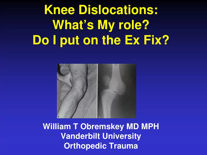
SLIDE 1
Knee Dislocations: What’s My role? Do I put on the Ex Fix?
William T Obremskey MD MPH Vanderbilt University Orthopedic Trauma
SLIDE 2 Disclosures
- Board SEFC
- OTA EBQVS Chair
- No Industry Conflicts
SLIDE 3 What’s My role?
– Reduce Joint – Assess Neuro/Vacular – Assist Vascular if needed – Release Compartments if needed – Stabilize - ?
SLIDE 4 Do I put on the Ex Fix?
SLIDE 5 OBJECTIVES
- What knee injuries are likely to result in
vascular injury?
- What is appropriate evaluation?
- When Ex Fix?
- Irreducible KD?
SLIDE 6
JAAOS December 2015
SLIDE 7 Schenck Classification KD I Multiligamentous injury with involvement of ACL or PCL KD II Injury to ACL and PCL only (2 ligaments) KD III Injury to ACL, PCL, and PMC or PLC (3 ligaments) KD IV Injury to ACL, PCL, PMC, and PLC (4 ligaments) KD V Multiligamentous injury with periarticular fracture
SLIDE 8 What Injuries?
What knee injuries are likely to result in vascular compromise?
- Fractures - distal femur and proximal
tibia
SLIDE 9
SLIDE 10
SLIDE 11
INJURY KNEE DISLOCATIONS
10% - 60% rate of associated vascular injury (5% - 15% requiring surgery)
SLIDE 12 High vs Low energy KNEE DISLOCATION
DeCoster JOT 1997 22 knee dislocations vs 28 “reduced” bicruciate ligament injuries
- 14% popliteal artery disruptions in
each
- Equal risk of vascular injury
SLIDE 13
VASCULAR INJURY TIMING
Miller Arch Surg 1949 Extremity salvage repair 90% at 6 hours 50% at 12-18 hours 20% at > 24 hours
SLIDE 14
DIAGNOSIS NONINVASIVE VASCULAR EXAM
Lynch, Johansen Ann Surg 1991 ABI < 0.9 95% sensitivity 97% specificity
SLIDE 15
SLIDE 16
When to Ex Fix?
SLIDE 17 When to Ex Fix?
– To manage CPS release
SLIDE 18 When to Ex Fix?
- Obese – unable to hold reduced
SLIDE 19 When to Ex Fix?
- Severe Soft Tissue injury
SLIDE 20
Nerve Injury
Peroneal most common
14% - 35%
One third will recover One half will remain as complete palsy
SLIDE 21
Peroneal Nerve Contusion
SLIDE 22
Nerve Avulsion
SLIDE 23 Indications for immediate
Open dislocation Irreducible dislocation Popliteal artery disruption Compartment syndrome
SLIDE 24
Open Dislocation
SLIDE 25
Open Dislocation Ex Fix or Splint
SLIDE 26
Posterolateral - irreducible
SLIDE 27
Irreducible
Pucker Sign
SLIDE 28
SLIDE 29
Evaluation: Radiographic Exam
AP/lat/oblique MRI helpful in defining torn structures Adds to both sensitivity and specificity (can still miss LCL and PLC) Aids preop planning by defining the location of tears
SLIDE 30 Take Home
Knee dislocation is challenging
Not always obvious When obvious, not always reducible closed
On table or formal Angiogram only for hard signs/ ABI < 0.9X Initial stabilization, then MRI prior to repair
SLIDE 31
THANK YOU