
SLIDE 1
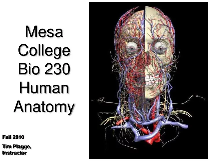
Mesa College Bio 230 Human Anatomy
Fall 2010 Tim Plagge, Instructor
Mesa College Bio 230 Human Anatomy Fall 2010 Tim Plagge, - - PowerPoint PPT Presentation
Mesa College Bio 230 Human Anatomy Fall 2010 Tim Plagge, Instructor Course Objectives Upon successful completion of this course you should: Know and be able to identify relevant tissues and microscopic structures of the human body
Fall 2010 Tim Plagge, Instructor
Wear open toed shoes in lab Have long hair that is not pulled back (so it doesn’t hang into specimens you may be working with) Eat or drink in lab Dissect the cadavers when they are out… that is another classes job.
You should:
Bring your book and any
hand outs that were given
Be prepared to use the
entire lab time
Bring gloves, or better yet,
keep a few pair in your book bag.
a. intervertebral cartilage b. cardiac muscle c. duodeno‐jujenum junction d.
Exams may consist of multiple choice, matching, true and false and short answer questions.
a.
b. dense irregular tissue c. dense irregular tissue d. hyaline cartilage Exams may consist of multiple choice, matching, true and false and short answer questions.
structures
– Microscopy (light & electron), CT scans, MRI, X‐rays, dissection . . . more later
– PET scans, ECG, sphygmomanometer . . .
– SEM (scanning electron microscopy) – TEM (transmission electron microscopy)
– Fluoroscope – x rays emitted through the specimen and images are viewed on a fluorescent screen – Cineradiography – uses X‐ray cinema film to record organ movements
Menu
Small/ Simple Large/ Complex
Figure 1.1
Developing neuron
– transport blood – regulate pressure & control volume
– carries O2 & CO2 – also carries nutrients & wastes – carries hormones – involved in hemostasis
– pumps blood – Creates pressure gradient for transportation and filtration
Menu
– Toward the upper part of a structure (or body), above
– Toward the lower part of a structure (or body), below
– Toward (or at) the front of the body (or structure)
– Toward (or at) the back of the body (or structure).
The heart is superior to the diaphragm The mouth is inferior to the nose The trachea is anterior to the esophagus The heart is posterior to the sternum
– Nearer or closer to the surface of the body, external
– Away or further from the body surface, internal
The epidermis is superficial to the dermis The muscles are deep to the skin
– Toward the midline of the body or structure
– Between a more medial and a more lateral structure
– Away from the midline of the body
– Closer to the origin of the body part or the point of attachment of a limb to the body trunk
– Further from the origin of the body part or the point of attachment of a limb to the body trunk
The sternum is medial to the scapula The nose is intermediate to,
The scapula are lateral to the vertebral column The shoulder is proximal to the elbow The ankle is distal to the knee
– Lies vertically and divides body into anterior and posterior parts
– Runs horizontally – divides body into superior and inferior parts
– Specific sagittal plane that lies vertically in the midline
Figure 1.11a, b
These regions are formed by two vertical planes and two horizontal planes.
The two vertical planes are the lateral lines LLL and RLL. These lines are dropped from a point half way between the jugular notch and the acromion process. The two horizontal planes are the transpyloric plane TPP and the transtubercular plane TTP. The tubercles are the tubercles of the iliac crests.
a pleural cavity
pericardial sac
and other organs
serosa (membrane) space in between = body cavity inner layer = visceral serosa (membrane)
Pericardial Cavity Pleural Cavity
Abdominal Cavity