Methods for determination of blood flow Stefan P. Mortensen, DMSc - - PDF document

methods for determination of blood flow
SMART_READER_LITE
LIVE PREVIEW

Methods for determination of blood flow Stefan P. Mortensen, DMSc - - PDF document

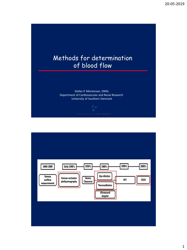
20-05-2019 Methods for determination of blood flow Stefan P. Mortensen, DMSc Department of Cardiovascular and Renal Research University of Southern Denmark 1 20-05-2019 Venous occlusion plethysmography 2 20-05-2019 Venous occlusion


slide-1
SLIDE 1

20-05-2019 1

Methods for determination

  • f blood flow

Stefan P. Mortensen, DMSc Department of Cardiovascular and Renal Research University of Southern Denmark

slide-2
SLIDE 2

20-05-2019 2

Venous occlusion plethysmography

slide-3
SLIDE 3

20-05-2019 3

Cuff

Venous occlusion plethysmography Venous occlusion plethysmography

slide-4
SLIDE 4

20-05-2019 4

Advantages:

  • Easy to use
  • Inexpensive
  • Non-invasive
  • Reliable

Disadvantages:

  • Exercise not possible

(early recovery)

  • Increase in venous pressure

can impede BF

  • Cuff pressure can impede

arterial blood flow

Venous occlusion plethysmography

50mmHg ~ 28% Pa Pv R

Q = (Pa – Pv) R

slide-5
SLIDE 5

20-05-2019 5

Pletysmography & xenon wash-out: Peak muscle blood flow = 50-60 ml/min/100g In pigs and other species: 200-400 ml/min/100g (Radiolabled microspheres)

Low peak blood flow compared to animals

Secher et al. 1977

Peak muscle blood flow = 50-60 ml/min/100 g 30 kg of muscle x 0.5 l/min = 15 l/min

slide-6
SLIDE 6

20-05-2019 6 . . .

  • +

Cuff

Strain-gauge

ring

  • M. quadriceps femoris
  • Art. femoralis
  • Ven. femoralis

biopsy

One legged knee-extensor model

Andersen and Saltin: J Physiol,1985

Constant infusion thermodilution

slide-7
SLIDE 7

20-05-2019 7

Chart Window

Blood temp. (degrees C) 34.5 35.0 35.5 36.0 36.5

  • Inf. temp (degrees C)

5 10 15 20 25 30 32:40 32:45 32:50 32:55 33:00 59 30-03-2006 15:06:29.251

1 ) ( ) ( ) ( ) (

1

−         − −             =

M B I B B B I I

T T T T C S C S V flow blood

Endurance trained athletes: 385 ml/min/100 g at 99 watts (Richardson et al. 1993) 15 kg x 4 l/min = 60 l/min (endurance trained subjects) Young subjects: 250 ml/min/100 g at 55 watts (Andersen & Saltin 1985) 15 kg x 2.5 l/min = 38 l/min (untrained subjects)

Peak perfusion rates

slide-8
SLIDE 8

20-05-2019 8

50 100 150 200 250 300 350 400 Low Moderate High Rat Peak BF Fitness ml/100g x min Advantages:

  • Can be used during whole-body exercise and maximal exercise
  • No recirculation
  • Easy to learn
  • Fairly inexpensive

Constant infusion thermodilution

Disadvantages:

  • Invasive
  • No resting measurements
  • Cooling

Temperature of surrounding tissue will be reduced after 15-18 s of infusion Tissue re-warming is slow (>45 s) Femoral arterial temperature is reduced after 20-25 s The ”problem” is exaggerated with high infusion rates and a low blood flow

  • Poor resolution
  • Haemodilution with many measurements
  • Some data interpretation
  • Availability
slide-9
SLIDE 9

20-05-2019 9

Dye dilution

Same principle as thermodilution, but with dye as the indicator instead of saline Arterial injection (bolus or continous) Venous detection photo-densitometer

slide-10
SLIDE 10

20-05-2019 10

Disadvantages:

  • Invasive
  • Expensive
  • Poor resolution
  • Allergic reactions to the dye can occur

Advantages:

  • Can be used during whole-body exercise and maximal exercise
  • Reliable measurements
  • Can be used to determine the mean transit time (MTT)
  • Can be combined with NIRS to determine regional blood flow

Dye dilution

slide-11
SLIDE 11

20-05-2019 11

Ultrasound Doppler

<60 (lower is better) Mean velocity Volume flow = Cross-sectional Area × Time-averaged velocity Area = π × radius2

Ultrasound Doppler

slide-12
SLIDE 12

20-05-2019 12

Advantages:

  • Can be used during exercise (forearm, leg kicking)
  • High resolution
  • Continuous measurement
  • Non-invasive

Disadvantages:

  • Sensitive to movement
  • Expensive equipment
  • Infusion noise
  • Difficult/time-consuming to learn

exercise measurements

Ultrasound Doppler Thermodilution versus Doppler

Rådegran 1997 Knee-extensor exercise ATP infusion

Thermodilution blood flow (l min-1) 1 2 3 4 5 Doppler blood flow (l min-1) 1 2 3 4 5 r=0.989

slide-13
SLIDE 13

20-05-2019 13

Microbubbles (SonoVue): 2.5 µm in diameter (range 0.7-10). SF6 gas encapsulated by a phospholipid shell Contrast-enhanced ultrasound (CEUS)

slide-14
SLIDE 14

20-05-2019 14

Contrast-enhanced ultrasound (CEUS) Contrast-enhanced ultrasound (CEUS)

Advantages:

  • Measures microcirculatory flow
  • Minimally invasive

Disadvantages:

  • Expensive
  • Difficult (impossible) to quantify
  • Difficult (impossible) to perform during execise – movement + contraction acts like a flash
  • Measures bubble flow – is it the same as plasma/erythrocyte flow?
slide-15
SLIDE 15

20-05-2019 15

Thank you for your attention!