
SLIDE 1
6/13/2019 1
N O F I N A N C I A L D I S C LO S U R E S
OBJECTIVES
Background When is it the right test? When is it the wrong test? What are some new things it might do?
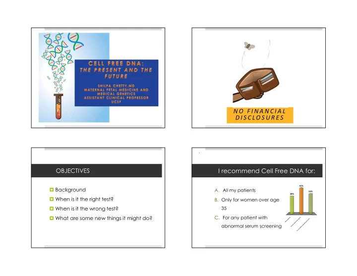
I recommend Cell Free DNA for:
- A. All my patients
- B. Only for women over age
35
- C. For any patient with
abnormal serum screening
A l l m y p a t i e n t s O n l y f
- r
w
- m
e n
- v
e r a g e 3 5 F
- r
a n y p a t i e n t w i t h a b n
- r
m a l s e r u m . . .
28% 32% 41%