Putting Putting T Tubes ubes W Within ithin T Tubes: ubes: - - PDF document

putting putting t tubes ubes w within ithin t tubes ubes
SMART_READER_LITE
LIVE PREVIEW

Putting Putting T Tubes ubes W Within ithin T Tubes: ubes: - - PDF document

12/11/2012 Disclosure Putting Putting T Tubes ubes W Within ithin T Tubes: ubes: Enteral Therapeutic Access Enteral Therapeutic Access Robert E. Kramer, MD Robert E. Kramer, MD Associate Professor of Pediatrics Associate Professor of


slide-1
SLIDE 1

12/11/2012 1

Putting Putting T Tubes ubes W Within ithin T Tubes: ubes: Enteral Therapeutic Access Enteral Therapeutic Access

Robert E. Kramer, MD Robert E. Kramer, MD

Associate Professor of Pediatrics Associate Professor of Pediatrics Director of Endoscopy Director of Endoscopy Digestive Health Institute Digestive Health Institute Children’s Hospital Children’s Hospital Colorado Colorado University University of Colorado

  • f Colorado

Disclosure

I have no financial relationships with any commercial entity to disclose

2

Objectives

  • Learn the various types of enteral

access including G, GJ, J and ceccal tubes/buttons

  • Recognize the indications and

appropriate usage for various access

  • ptions
  • Know proper placement and care

techniques to minimize complications

Background

  • Wide variety of indications for enteral tube

placement in children

  • Determination of most appropriate device for is

dependent on

  • Indication
  • Anticipated duration
  • Need for fundoplication
  • Current feeding device
  • Anatomic considerations

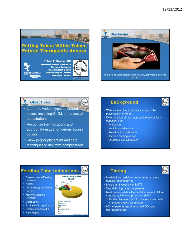
Feeding Tube Indications

  • Developmental Feeding

problems

  • Allergy
  • Inflammatory conditions
  • Surgery

6% 3% 3% 2% 1% 6%

Indications for PEG N=239

  • Surgery
  • Motility Disorders
  • HIV/AIDS
  • Short Bowel
  • Aspiration/ Lung disease
  • Chronic disease c FTT
  • Pancreatitis

79%

Neuro Impairment Myopathy Dysphagia CF Metabolic D/O HIV Misc

Fiscetti-Leon F, Dig Liv Dis, 2012

Timing

  • No definitive guidelines for transition to more

durable feeding device

  • More than 8 weeks with NGT?
  • Very difficult process for parents
  • Most parents of developmentally delayed children

very happy following procedure (91%)

  • Earlier placement (< 18 mos) associated with

improved growth parameters

  • 85% of parents report improved QOL and

decreased stress

slide-2
SLIDE 2

12/11/2012 2

Feeding Tube Options

  • Nasoenteral
  • NGT
  • NJT
  • Gastrostomy
  • PEG/endoscopic
  • Surgical
  • Radiologic
  • Transpyloric/ Gastrojejunostomy
  • Initial placement
  • Conversion from existing gastrostomy
  • Enterostomy
  • PEJ
  • Surgical jejunostomy
  • cecostomy

7 7

Feeding Spectrum

8

Least Invasive Most Invasive

Nasogastric/Nasojejunal

Pros

  • Easy, most temporary
  • Typically placed under

fluoroscopic guidance

  • Endo placement for

Cons

  • Invasive to replace
  • Uncomfortable
  • Easily removed
  • Easily displaced by

difficult anatomy or when diagnostic endoscopy needed

  • Easily removed
  • May use as trial

vomiting

  • Long term Complications
  • Sinusitis
  • Esophageal/gastric

erosions

  • Fatal hemorrhage from

aorto-esophageal fistulae

Methods: NDT Placement

  • Primarily placed by Radiology under fluoroscopy
  • Endo placement due to pt size or altered anatomy
  • Generally use “drag method”
  • Pitfall of drag method is removal of scope from

Pitfall of drag method is removal of scope from duodenum without displacement of tube

  • Polyp snare vs clip method
  • Clip method: create suture loop at tip
  • Caution: loop tangling with clip

Polyp-Snare Method of NDT Placement

11

Endoclip Method of NDT Placement

12

slide-3
SLIDE 3

12/11/2012 3

  • Placed by GI
  • Avoids surgical incision
  • Shorter recovery time (d/c

within 1-2 days) Cons

  • Risk of perforation (2-3%)
  • No antireflux protection
  • Most Neurologically impaired

children, even with significant reflux, do well even w/o Nissen

PEG vs Surgical Gastrostomy

Pros

  • Less invasive, better

tolerated by critically ill pts

  • Able to use later that day
  • Decreased medical costs
  • Complication rate

comparable to surgical method (19% vs 11%)

Nissen

  • May increase risk of reflux
  • Long tube, needs to be converted

to button device

  • Contraindicated if altered

anatomy

  • severe scoliosis
  • malrotation
  • ? Prior abdominal surgery

PEG Placement: "Scoping” Side

14

PEG Placement: “Poking” Side

15

Post-PEG Care

  • Cefazolin 20 mg/kg IV intraop, 6 hrs postop
  • NPO x 4-6 hrs, then Pedialyte 60 cc bolus
  • Can take bath after 7 days
  • May swim after 2 weeks
  • Clean and rotate tube 180° 1-2 times per day
  • Flush tube with 15 ml of water after each use
  • May try Club soda if clogged
  • May still have “tummy time”, foam donut if irritated
  • Change to button 8-12 weeks after PEG placement
  • Pull method versus endoscopic
  • Inadvertent removal before 6 weeks, confirm placement with film
  • Granulation tissue: triamcinalone 0.5%, silver nitrate

16

Gastrostomy Tube Types

17

MIC-Key Button BARD Skin Level Corpak Corflo Cubby AMT Mini One Kendall NutriPort

Gastrojejunostomy

  • Conversion from GT can usually be done by

Radiology (wt >10 kg)

  • Endoscopically, easiest to pass scope through

stoma (XP180, 5.6 mm, 16 Fr) and thread wire through scope and then tube over wire.

  • Must choose appropriate length GJ. Too long and

tube tends to loop in stomach from back-pressure.

  • Angle scope toward pylorus
  • Use Murray-lube and stiff guidewire
  • Schedule replacement every 3 months by

Radiology

  • Easier than starting from scratch if becomes dislodged
slide-4
SLIDE 4

12/11/2012 4

Initial Percutaneous GJ Placement

  • 1. Stay sutures to anchor stomach
  • 2. Angle insertion toward pylorus
  • 3. Avoid driving dilators into opposing wall
  • 4. Pass guidewire AFTER dilation

19 19

Jejunostomy

  • Indications: Direct access to small bowel,

repeated loss of GJ placement

  • Methods: PEJ vs Surgical
  • Consider PEJ:
  • Smaller children (< 20 kg) when balloon may obstruct lumen

( g) y

  • When more invasive surgery difficult to tolerate
  • History of multiple GI surgeries
  • Technique:
  • Same premise as PEG, but anatomy not as defined
  • May do hybrid Lap-assisted PEJ with surgeon
  • Published literature: Small series of 5 patients, with 2

minor complications

20

Cecostomy

  • Indications: severe constipation,

refractory to medical therapy

  • Anorectal malformation, Hirschsprung’s, CP, idiopathic, spina bifida
  • Methods: Surgical, percutaneous non-endoscopic,

d i (PEC) l i i t d (LAPEC) endoscopic (PEC), laparoscopic assisted (LAPEC)

  • Technique
  • Similar to PEG technique
  • With LAPEC use single umbilical port to assist passage of scope and

stabilization/visualization of cecum during trochar placement

  • Complications: overall 16-30%
  • Chait: 6% site infection, 10% tube failure, 14% tube dislodgement
  • LAPEC: 2% hematoma, 12% fever, 6% dislodgement, 4% skin erosion

21

Take-Home Points

  • Variety of techniques available for endoscopic

placement of enteral devices

  • Complicated psychosocial aspects surrounding

placement

  • Choice of most appropriate device/technique

depends on indication, anatomy, anticipated duration and size of patient

  • Significant risks for placement, comparable to

surgical placement

Future Directions

  • Larger, randomized trials needed to compare

surgical, endoscopic and radiologic methods for enteral access

  • Development of hybrid laparoscopic/endoscopic

p y p p p procedures to minimize invasiveness and costs while maximizing safety

  • Application of principles of Natural Orifice

Transluminal Endoscopic Surgery (NOTES) to process of enteral device placement

23

References

  • 1. Goretsky MF, Alternative techniques of feeding gastrostomy in children: a critical
  • analysis. J Am Coll Surg, 1996. PMID 8603243
  • 2. Martinez-Costa C. Early decision of gastrostomy tube insertion in children with severe

developmental disability: a current dilemma. J Hum Nutr Diet, 2011. PMID 21332837

  • 3. Kawahara H. Should fundoplication be added at the time of gastrostomy placement in

ti t h l i ll i i d? J P di t S 2010 PMID 21129548 patients who are neurologically impaired? J Pediatr Surg 2010. PMID 21129548

  • 4. Miura T. A Fatal Aortoesophageal Fistula Caused by Critical Combination of Double

Aortic Arch and Nasogastric Tube Insertion for Superior Mesenteric Artery Syndrome. Case Reports Gastroenterol 2010. PMID 20805944

  • 5. Minar P. Safety of percutaneous endoscopic gastrostomy in medically complicated
  • infants. J Pediatr Gastroenterol Nutr 2011. PMID 21865977
  • 6. Toporowska-Kowalska E. Influence of percutaneous endoscopic gastrostomy on gastro-
  • esophageal reflux evaluated by multiple intraluminal impedance in children with

neurological impairment. Dev Med child Neuro 2011. PMID 21752017

  • 7. Faqundes RB. Percutaneous endoscopic gastrostomy and peristomal infection: an

avoidable complication with the use of a minimum skin incision. Surg Laparosc Endosc Percutan Tech 2011. PMID 21857479

slide-5
SLIDE 5

12/11/2012 5

References (Continued)

  • 8. Schie CB. Clip-assisted endoscopic method for placement of a nasoenteric feeding tube

into the distal duodenum. J Formos Med Assoc. 2003. PMID 14517593

  • 9. Avitsland TL, et al. Maternal psychological distress and parenting stress after

gastrostomy placement in children. JPGN, epub May 2012. PMID 22644463

  • 10 Fascetti-Leon F et al Complications of percutaneous endoscopic gastrostomy in
  • 10. Fascetti Leon F, et al. Complications of percutaneous endoscopic gastrostomy in

children: Results of Italian multicenter observational study. Dig Liv Dis, 2012; 44(8):655-9. PMID 22541388

  • 11. Virnig DJ, et al. Direct percutaneous endoscopic jejunostomy: a case series in pediatric
  • patients. Gastrointest Endosc 2008; 67(6): 984-87. PMID 18308316
  • 12. Rodriguez L, et al. Laparoscopic-assisted percutaneous endoscopic cecostomy in

children with defecation disorders (with video). Gastrointest Endosc 2011; 73(1):98-102 PMID 21184875

  • 13. Chait PG, et al. Percutaneous cecostomy: updates in technique and patient care.

Radiology 2003;227:246-50.

25