Table of Contents Why DNA Computing? The Structure of DNA DNA - - PDF document

table of contents
SMART_READER_LITE
LIVE PREVIEW

Table of Contents Why DNA Computing? The Structure of DNA DNA - - PDF document

Table of Contents Why DNA Computing? The Structure of DNA DNA Computing Operations on DNA Molecules Reading DNA Example of a Molecular Computer Information Processing with DNA Molecules Christian Jacob Why DNA Computing?


slide-1
SLIDE 1

DNA Computing

Information Processing with DNA Molecules

Christian Jacob

Table of Contents

Æ Why DNA Computing? Æ The Structure of DNA Æ Operations on DNA Molecules Æ Reading DNA Æ Example of a Molecular Computer

Why DNA Computing?

Æ From silico to carbon.

From microchips to DNA molecules.

Æ Limits to miniaturization with current

computer technologies.

Æ Information processing capabilities of

  • rganic molecules ...

Æ replace digital switching primitives, Æ enable new computing paradigms.

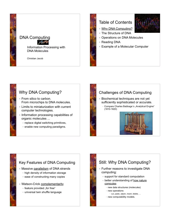
Challenges of DNA Computing

Æ Biochemical techniques are not yet

sufficiently sophisticated or accurate.

Æ Compare Charles Babbage´s „Analytical Engine“

(1810-1820)

Key Features of DNA Computing

Æ Massive parallelism of DNA strands

Æ high density of information storage Æ ease of constructing many copies

Æ Watson-Crick complementarity

Æ feature provided „for free“ Æ universal twin shuffle language

Still: Why DNA Computing?

Æ Further reasons to investigate DNA

computing:

Æ support for standard computation Æ better understanding of how nature

computes

Æ new data structures (molecules) Æ new operations

l cut, paste, adjoin, insert, delete, ...

Æ new computability models.

slide-2
SLIDE 2

Table of Contents

Æ Why DNA Computing? Æ The Structure of DNA Æ Operations on DNA Molecules Æ Reading DNA Æ Example of a Molecular Computer

The Structure of DNA

Æ DNA is a polymer („large“ molecule). Æ DNA is strung together from monomers

(„small“ mols.): deoxyribonucleotides.

Æ DNA = Deoxyribo Nucleic Acid Æ DNA supports two key functions for life:

Æ coding for the production of proteins, Æ self-replication.

Structure of a DNA Monomer

Æ Each deoxyribonucleotide consists of

three components:

Æ a sugar — deoxyribose

Æ five carbon atoms: 1´ to 5´ Æ hydroxyl group (OH)

attached to 3´ carbon

Æ a phosphate group Æ a nitrogenous base.

Chemical Structure of a Nucleotide

Structure of a DNA Monomer (2)

Æ DNA nucleotides differ only by their

bases (B):

Æ purines

Æ Adenine

A

Æ Guanine

G

Æ pyrimidines

Æ Cytosine

C

Æ Thymine

T

Linking of Nucleotides

Æ The DNA monomers can link in two ways:

Æ Hydrogen bond Æ Phosphodiester bond

slide-3
SLIDE 3

Linking of Nucleotides

Phosphodiester Bond

Æ The 5´-phosphate group of one nucleotide is

joined with the 3´-hydroxyl group of the other

Æ strong (covalent) bond Æ directionality:

5´—3´ or 3´—5´

Linking of Nucleotides

Phosphodiester Bond

Linking of Nucleotides

Hydrogen Bond

Æ The base of one nucleotide interacts with the

base of another

Æ base pairing (weak bond)

l A — T

(2 hydrogen bonds)

l C — G

(3 hydrogen bonds)

Æ Watson-Crick complementarity

l James D. Watson l Francis H. C. Crick l deduced double-helix

structure of DNA in 1953

l Nobel Prize (1962)

Linking of Nucleotides

Hydrogen Bond

DNA

Double Helix

Æ Longer streches keep the double

strands together through the cumulative effect (the sum) of hydrogen bonds.

Æ Dense packing:

l Bacteria: DNA molecule is 10,000 times

longer than the host cell

l Eucaryotes:

„hierarchical“ packing

Table of Contents

Æ Why DNA Computing? Æ The Structure of DNA Æ Operations on DNA Molecules Æ Reading DNA Æ Example of a Molecular Computer

slide-4
SLIDE 4

Operations on DNA Molecules

Æ Separating and fusing DNA strands Æ Lengthening of DNA Æ Shortening DNA Æ Cutting DNA Æ Multiplying DNA

Separating and Fusing DNA Strands

Æ Denaturation: separating the single

strands without breaking them

Æ weaker hydrogen than phosphodiester

bonding

Æ heat DNA (85° - 90° C)

Æ Renaturation:

Æ slowly cooling down Æ annealing of matching, separated strands

Enzymes

Machinery for Nucleotide Manipulation

Æ Enzymes are proteins that catalyze

chemical reactions.

Æ Enzymes speed up chemical reactions

extremely efficiently (speedup: 1012)

Æ Enzymes are very specific. Æ Nature has created a multitude of

enzymes that are useful in processing DNA.

Lengthening DNA

Æ DNA polymerase enzymes

add nucleotides to a DNA molecule

Æ Requirements:

Æ single-stranded template Æ primer,

l bonded to the template l 3´-hydroxyl end available for

extension

l Note: Terminal transferase

needs no primer.

Shortening DNA

Æ DNA nucleases are enzymes

that degrade DNA.

Æ DNA exonucleases

l cleave (remove) nucleotides one at

a time from the ends of the strands

l Example: Exonuclease III

3´-nuclease degrading in 3´-5´direction

Shortening DNA

Æ DNA nucleases are enzymes

that degrade DNA.

Æ DNA exonucleases

l cleave (remove) nucleotides one at

a time from the ends of the strands

l Example: Bal31

removes nucleotides from both strands

slide-5
SLIDE 5

Cutting DNA

Æ DNA nucleases are enzymes

that degrade DNA.

Æ DNA endonucleases

l destroy internal phosphodiester

bonds

l Example: S1

cuts only single strands or within single strand sections

Æ Restriction endonucleases

l much more specific l cut only double strands l at a specific set of sites (EcoRI)

Multiplying DNA

Æ Amplification of a „small“ amount of a specific

DNA fragment, lost in a huge amount of other pieces.

Æ „Needle in a haystack“ Æ Solution: PCR = Polymerase Chain Reaction

Æ devised by Karl Mullis in 1985 Æ Nobel Prize Æ a very efficient molecular Xerox machine

PCR

Step 0: Initialization

Æ Start with a solution containing

the following ingredients:

l the target DNA molecule l primers

(synthetic oligonucleotides), complementary to the terminal sections

l polymerase,

heat resistant

l nucleotides

PCR

Step 1: Denaturation

Æ Solution heated close to boiling

temperature.

Æ Hydrogen bonds between the

double strands are separated into single strand molecules.

PCR

Step 2: Priming

Æ The solution is cooled down (to

about 55° C).

Æ Primers anneal to their

complementary borders.

PCR

Step 3: Extension

Æ The solution is heated again (to

about 72° C).

Æ A polymerase will extend the

primers, using nucleotides available in the solution.

Æ Two complete strands of the

target DNA molecule are produced.

slide-6
SLIDE 6

PCR

Efficient Xeroxing: 2n copies after n steps

Step 1 Step 2 Step 3 Step 4 Step 5

Table of Contents

Æ Why DNA Computing? Æ The Structure of DNA Æ Operations on DNA Molecules Æ Reading DNA Æ Example of a Molecular Computer

Measuring the Length of DNA Molecules

Gel Electrophoresis

Æ DNA molecules are negatively charged. Æ Placed in an electric field, they will move

towards the positive electrode.

Æ The negative charge is proportional to the length

  • f the DNA molecule.

Æ The force needed to move the molecule is

proportional to its length.

Æ A gel makes the molecules move at different

speeds.

Æ DNA molecules are invisible, and must be

marked (ethidium bromide, radioactive)

Schematic representation

  • f gel electrophoresis

Radioactive marker Ethidium bromide marker

Sequencing a DNA Molecule

Æ Sequencing:

Æ reading the exact sequence of nucleotides

comprising a given DNA molecule

Æ based on

l the polymerase action of extending a primed single

stranded template

l nucleotide analogues l chemically modified l e.g., replace 3´-hydroxyl group (3´-OH) by 3´-

hydrogen atom (3´-H)

l dideoxynucleotides:

  • ddA, ddT, ddC, ddG

l Sanger method, dideoxy enzymatic method

Sequencing — Part 1

Æ Objective

Æ We want to sequence a single stranded molecule

.

Æ Preparation

Æ We extend at the 3´ end by a short (20 bp)

sequence , which will act as the W-C complement for the primer compl().

l Usually, the primer is labelled (radioactively, or marked

fluorescently)

Æ This results in a molecule ´= 3´- .

slide-7
SLIDE 7

Sequencing — Part 2

Æ 4 tubes are prepared:

l Tube A, Tube T, Tube C, Tube G l Each of them contains l molecules l primers, compl() l polymerase l nucleotides A, T, C, and G. l Tube A contains a limited amount of ddA. l Tube T contains a limited amount of ddT. l Tube C contains a limited amount of ddC. l Tube G contains a limited amount of ddG.

Reaction in Tube A

Æ The polymerase enzyme

extends the primer of ´, using the nucleotides present in Tube A: ddA, A, T, C, G.

Æ using only A, T, C, G:

l ´ is extended to the full duplex.

Æ using ddA rather than A:

l complementing will end at the

position of the ddA nucleotide.

Resulting Sequences in Tubes

Æ Tube A:

Æ TCATGCACTGCG Æ TCA Æ TCATGCA

Æ Tube T:

Æ TCATGCACTGCG Æ T Æ TCAT Æ TCATGCACT

Æ Tube C:

Æ TCATGCACTGCG Æ TC Æ TCATGC Æ TCATGCAC Æ TCATGCACTGC

Æ Tube G:

Æ TCATGCACTGCG Æ TCATG Æ TCATGCACTG

Final Reading of the Strands

Æ Tube A: l

TCATGCACTGCG

l

TCA

l

TCATGCA

Æ Tube T: l

TCATGCACTGCG

l

T

l

TCAT

l

TCATGCACT

Æ Tube C:

l TCATGCACTGCG l TC l TCATGC l TCATGCAC l TCATGCACTGC

Æ Tube G:

l TCATGCACTGCG l TCATG l TCATGCACTG

Gel electrophoresis:

Æ We read:

Æ T Æ TC Æ TCA Æ TCAT Æ TCATG Æ TCATGC Æ TCATGCA Æ TCATGCAC Æ TCATGCACT Æ TCATGCACTG Æ TCATGCACTGC Æ TCATGCACTGCG

Table of Contents

Æ Why DNA Computing? Æ The Structure of DNA Æ Operations on DNA Molecules Æ Reading DNA Æ Example of a Molecular Computer

Adleman´s Experiment

Æ In 1994 Leonard M. Adleman showed how to

solve the Hamilton Path Problem, using DNA computation.

Æ Hamiltonian Path Problem:

Æ Let G be a directed graph with designated input

and output vertices, vin and vout.

Æ Find a (Hamiltonian) path from vin to vout that

involves every vertex in G exactly once.

slide-8
SLIDE 8

vin vout

2 5 6 1 4 3

Hamiltonian Path Example

Æ Adleman´s graph Æ The only Hamiltonian path

for this graph is:

Æ 0—1—2—3—4—5—6

Æ Simplified graph Æ Hamiltonian path:

l Atlanta l Boston l Chicago l Detroit

vin vout

Adleman´s Algorithm

Æ Input: A directed graph G with n vertices, and

designated vertices vin and vout.

Æ Step 1: Generate paths in G randomly in large

quantities.

Æ Step 2: Reject all paths that

l do not begin with vin and l do not end in vout.

Æ Step 3: Reject all paths that do not involve exactly n

vertices.

Æ Step 4: For each of the n vertices v:

l reject all paths that do not involve v.

Æ Output: YES, if any path remains; NO, otherwise.

Vertex and Edge Encodings

Æ Each city vi is encoded by two sub-sequences: vi = vi´

vi´´ Each flight eik from vi to vk is encoded by: eik = vi´´ vk´

Town DNA Name Complement Flight DNA Flight Number

DNA Computation

Æ The town complements and DNA flight numbers are used for

computation.

Æ DNA molecules are put in a hydrous solution. Æ Addition of ligase ensures catalysis of phosphodiester bonds. Æ Shaking the test tube makes many DNA strands collide and

interact.

Æ ~1014 computations are carried out in a single second. Æ The solution strand has to be filtered from the test tube:

l GCAG TCGG ACTG GGCT ATGT CCGA l Atlanta Boston Chicago Detroit

„DNA Computer“

Performance Evaluation

Æ Information density: l 1015 CDs per cm3 Æ Massively parallel information processing:

l 106 ops / sec for PCs l 1012 ops / sec for supercomputers l 1020 ops / sec possible for DNA l DNA computers would be > 1,000,000 times faster than any

computer today.

Æ Energy efficiency: l 2 * 1019 operations per joule for DNA l 109 operations/joule for silicon-based computers

Table of Contents

Æ Why DNA Computing? Æ The Structure of DNA Æ Operations on DNA Molecules Æ Reading DNA Æ Example of a Molecular Computer

slide-9
SLIDE 9

References

Æ Paun, G., Rozenberg, G., and Salomaa,

A., DNA Computing, Springer,1998.