
SLIDE 1
5/3/2018 1
The Red Eye
John Knapp, MD
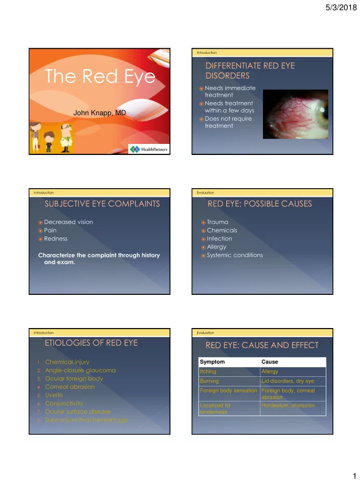
⦿ Needs immediate
treatment
⦿ Needs treatment
within a few days
⦿ Does not require
treatment
Introduction
DIFFERENTIATE RED EYE DISORDERS SUBJECTIVE EYE COMPLAINTS
⦿ Decreased vision ⦿ Pain ⦿ Redness
Characterize the complaint through history and exam.
Introduction
RED EYE: POSSIBLE CAUSES
⦿ Trauma ⦿ Chemicals ⦿ Infection ⦿ Allergy ⦿ Systemic conditions
Evaluation
ETIOLOGIES OF RED EYE
- 1. Chemical injury
- 2. Angle-closure glaucoma
- 3. Ocular foreign body
- 4. Corneal abrasion
- 5. Uveitis
- 6. Conjunctivitis
- 7. Ocular surface disease
- 8. Subconjunctival hemorrhage
Introduction
RED EYE: CAUSE AND EFFECT
Symptom Cause Itching Allergy Burning Lid disorders, dry eye Foreign body sensation Foreign body, corneal abrasion Localized lid tenderness Hordeolum, chalazion
Evaluation