SLIDE 2 Journal of Islamabad Medical & Dental College (JIMDC); 2015:4(4):176-177 177
There was no power deficit. Rest of the neurological examination was normal. MRI Cervical Spine showed hyperintense lesion at level of C3, 4, 5, 6 on T1 image and homogenously enhancing with gadolinium contrast as shown in Figure.1. The Patient was
- perated. The tumor was adherent but was separable
from the dura and total excision was obtained. Histology showed meningioma with EMA positive on
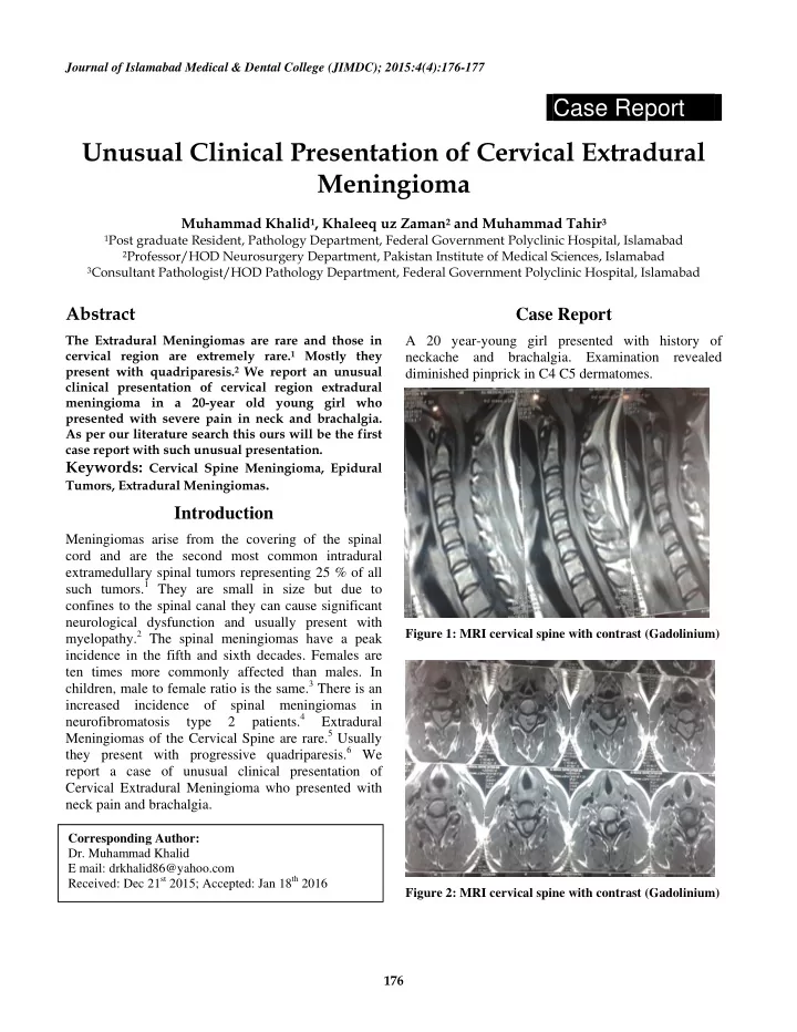
- Immunohistochemistry. Axial cuts with contrast
showed extradural contrast enhancing lesion extending into C4/5 neural foramen along the nerve root as shown in Figure 2.
Discussion
Among the intraspinal tumors, meningiomas are the second most common tumors. It is four times more common in women than men and usually occurs in the fifth to sixth decades of life. Almost 80% of these tumors arise from the thoracic spine.7 These spinal meningiomas are intradural extramedullary lesions and are mostly ventrally or ventro-laterally placed.8 There is an extradural component in almost 10% of the cases but it is rare to have an exclusively extradural meningioma.9 It is still unclear how meningiomas arise from extradural site and one of the possibility is due to abnormally located meningothelial cells in this region.10 Mostly extradural spinal lesions are metastatic neoplasms and can also be due to lymphomas and therefore it is essential to make a correct diagnosis and to exclude these extradural lesions to have a proper treatment plan accordingly.11 In patients having a younger age group and in whom there is a negative metastatic evaluation, other possibilities should be considered such as meningiomas, neurofibromas, schwannomas, chordomas, infectious lesions or synovial cysts.12 In the literature reviewed there were 17 case reports of extradural meningioma found. Most of the patients presenting with extradural meningioma were of 14 to 75 years age group, and among these 47% of the patients were younger than 30 years of age. In most of the cases the affected individuals were females (64.7%) similar results were seen in our case. Among the female patients 75% of cases were younger than 30 years of age. This predilection of age along with gender may be due to association of hormonal effects
- n the growth and development of meningioma.
Among various studies, in 52.9% of cases reviewed, the most common site involved was thoracic region and in 41.2% of the cases cervical extradural meningiomas were present.13 Usually these meningiomas present with progressive quadriparesis and no case of cervical extradural meningioma presenting with neck-ache and brachalgia has been reported.6
Conclusion
Cervical extradural meningioma can present in unusual pattern both clinically and radiologically. It should be kept in mind in patients presenting with neck-ache and brachalgia.
References
- 1. Osborn AG. Diagnostic neuroradiology. Mosby
Inc.1994; ISBN:0801674867.
- 2. Soderlund KA, Smith AB, Rushing EJ et-ai. Radiologic-
pathologic correlation of pediatric and adolescent spinal neoplasms: part 2, intradural extramedullary spinal neoplasms. AJR Am J Roentgenol.2012;198(1): 44-51
- 3. Buetow MP, Buetow PC, Smirniotopoulos JG. Typical,
Atypical, and misleading features in meningioma. Radiographics 1991;11(6):1086-1106.
- 4. Abul-kasim K, thurnher MM, Mckeever P et-al.
Intradural spinal tumors: current classification and MRI features. Neuroradiology.2008;50(4):301-14.
- 5. Brian L Frank, James S Harrop, Amgad Hanna, John
- Ratliff. Cervical Extradural Meningioma: Case Report
and Literature Review. J Spinal Cord Med 2008;31(3): 302–305.
- 6. Takeuchi H, Kubota T, Sato K, Hirose S. Cervical
extradural meningioma with rapidly progressive
- myelopathy. Journal of Clinical Neuroscience April
2006;13(3): 397–400.
- 7. Tuli J, Drzymalski DM, Lidov H, Tuli S. Extradural
en‑ plaque spinal meningioma with intraneural
- invasion. World Neurosurg 2012; 77:202.e5‑ 13.
- 8. Santiago BM, Rodeia P, Cunha E Sa M. Extradural
thoracic spinal meningioma. Neurol India 2009; 57:98.
- 9. Zevgaridis D, Thomé C. Purely epidural spinal
meningioma mimicking metastatic tumor: Case report and review of literature. Spine (Phila Pa 1976) 2002;27:E403‑ 5.
M, Abu Eid M, Bogorin A et al., “Lesm´eningiomes rachidiens extraduraux: donn´ees IRM ´a propos de deux observations,” Journal of Neuroradiology, vol. 31, no. 3, pp. 214–219, 2004.
- 11. Savardekar A, Chatterjee D, Chatterjee D, Dhandapani
S, Mohindra S, Salunke P. Totally extradural spinal en plaque meningiomas - Diagnostic dilemmas and treatment strategies. Surg Neurol Int 2014;5: S291-4.
- 12. Ross J, Brant-Zawadzki M, Chen M, Moore K, Salzman
- K. Diagnostic Imaging: Spine. 1st ed. Salt Lake City,
UT: Amirsys; 2004. Meningioma; IV1–78-IV1-81.