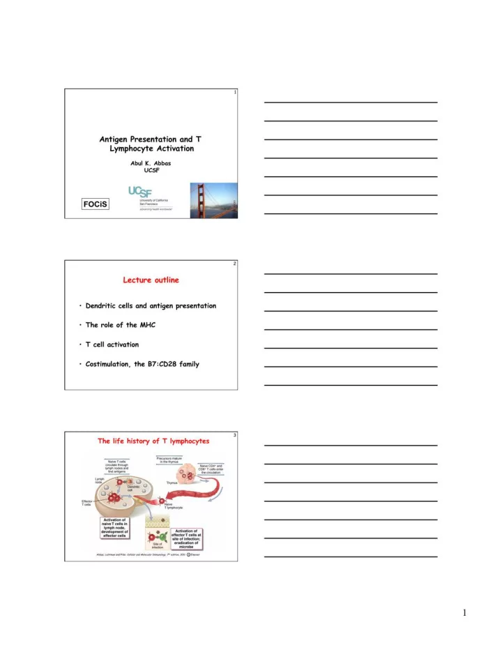
2 10 Why are dendritic cells the most efficient APCs for - - PDF document
4 The challenge for lymphocytes Very few lymphocytes in the body are specific for any one microbe (or antigen) Specificity and diversity of antigen receptors: the immune system recognizes and distinguishes between 10 6 - 10 9 antigens;
2
- 4
The challenge for lymphocytes
- Very few lymphocytes in the body are
specific for any one microbe (or antigen)
– Specificity and diversity of antigen receptors: the immune system recognizes and distinguishes between 106 - 109 antigens; therefore, few lymphocytes with the same receptors
5
The challenge for lymphocytes
- Very few lymphocytes in the body are specific for any one microbe
(or antigen) – Specificity and diversity of antigen receptors: the immune system recognizes and distinguishes between 106 - 109 antigens
- Lymphocytes must be able to locate
microbes that enter and reside anywhere in the body
– Usual routes of entry are through epithelia, but infections may take hold anywhere
6
The challenge for lymphocytes
- Very few lymphocytes in the body are specific for any one microbe
(or antigen)
- Lymphocytes must be able to locate microbes that enter anywhere in
the body
- Lymphocytes must respond to each microbe
in ways that are able to eradicate that microbe; best exemplified by T cells
– Extracellular microbes: antibodies; destruction in phagocytes (need helper T cells) – Intracellular microbes: killing of infected cells (need CTLs) – How do T cells distinguish antigens in different cellular locations?
4
- 10
Why are dendritic cells the most efficient APCs for initiating immune responses?
- Location: at sites of microbe entry (epithelia), tissues
- Receptors for capturing and reacting to
microbes: Toll-like receptors, other receptors
- Migration to T cell zones of lymphoid organs
– Role of CCR7 – Co-localize with naïve T cells
- Maturation during migration: Conversion from cells
designed for antigen capture into cells for antigen presentation and T cell activation
- Practical application: dendritic cell-based vaccines
for tumors
Take home messages
11
What do T cells see?
- All functions of T cells are mediated by
interactions with other cells
– Helper T cells “help” B cells to make antibodies and “help” macrophages to destroy what they have eaten – Cytotoxic (killer) T lymphocytes kill infected cells
- How does the immune system ensure that
T cells see only antigens on other cells?
12
What do T cells see?
- All functions of T cells are mediated by
interactions with other cells
– Helper T cells “help” B cells to make antibodies and “help” macrophages to destroy what they have eaten – Cytotoxic (killer) T lymphocytes kill infected cells
- To ensure cellular communications, T cells
see antigens NOT in the circulation but
- nly when displayed by molecules on the
surface of other cells
– These molecules are HLA (generic name: MHC) and the cells displaying the antigen are APCs
Take home messages
6
- 16
- Macrophage
ingests microbe Microbial antigen is presented by class II MHC Antigen is recognized by CD4+ T cell CD4+ T cells secrete cytokines that activate macrophage to destroy ingested microbe B cell recognizes antigen and ingests it Antigen is presented by class II MHC Antigen is recognized by CD4+ T cell CD4+ T cells secrete cytokines that activate B cells to differentiate into antibody secreting plasma cells
Functional importance of class II MHC- associated antigen presentation
17
- Viral antigen is
produced inside virus-infected cell Viral antigen is presented by class I MHC Antigen is recognized by CD8+ cytotoxic (killer) T cell Infected cell is killed, eliminating the infection
Functional importance of class I MHC- associated antigen presentation
18
Functions of antigen-presenting cells
- Capture antigens and take them to the
“correct” place
– Antigens are concentrated in peripheral lymphoid organs, through which naïve lymphocytes circulate
- Display antigens in a form that can be
recognized by specific lymphocytes
– For T cells: MHC-associated peptides (cytosolic peptides to class I, vesicular peptides to class II) – For B cells: native antigens
- Provide “second signals” for T cell
activation
– Critical for initiation of responses
Take home messages
10
- 28
- Major functions of selected B7-CD28
family members
- B7-CD28: initiation of immune
responses
- ICOS-ICOS-L: T cell help in
germinal center reactions (antibody responses)
- B7-CTLA-4: inhibits early T cell
responses in lymphoid organs
- PD-1:PD-L1,2: inhibits effector T
cell responses in peripheral tissues
Activation Inhibition
29
- Complexities and unknowns of B7:CD28
costimulation
- Different T cell populations vary in their
dependence on B7:CD28:
– Naïve > activated > memory – CD4 > CD8 – Regulatory T cells (controllers of immune responses) are also B7-dependent
- Redundancy of B7-1 and B7-2?
- Does B7 signal backwards into APCs?
30
- Therapeutics based on the B7:CD28 family
- 1. Costimulatory blockade
CTLA-4.Ig inhibits T cell activation in diseases caused by T cell responses-- rheumatoid arthritis, graft rejection, psoriasis
11
- 31
- Costimulatory blockade therapy
- B7-antagonist (CTLA-4.Ig, Abatacept)
approved for RA, kidney allograft rejection (high-affinity version, Belatacept) – Risks:
- Reducing responses against infections
32
- 32
- Costimulators other than B7:CD28
- Many proteins of the TNF-receptor
family are expressed on T cells and implicated in T-cell activation and control
– Functions often demonstrated in complex experimental systems or in vitro – Roles in disease (human or animal models) not definitely established
- Possible therapeutic targets?
33
- T cell expansion and contraction (decline)
Many aspects of T cell responses and functions are mediated by cytokines: initial activation -- IL-2; maintenance of memory cells -- IL-7; effector functions -- various
12
- Clonal expansion of T cells
- Stimulated mainly by autocrine IL-2
– Antigen recognition secretion of IL-2 and expression of high-affinity IL-2 receptors preferential expansion of antigen-specific cells
- CD8+ T cells may expand >50,000-fold
within a week after an acute viral infection
– Up to 10% of all CD8+ T cells in the blood may be specific for a pathogen – Minimal expansion of “bystander” cells (not specific for the virus) – CD8+ cells expand much more than do CD4+ cells
34