
SLIDE 1
Pathogens and commensals: War and Peace at mucosal surface Nothing - - PowerPoint PPT Presentation
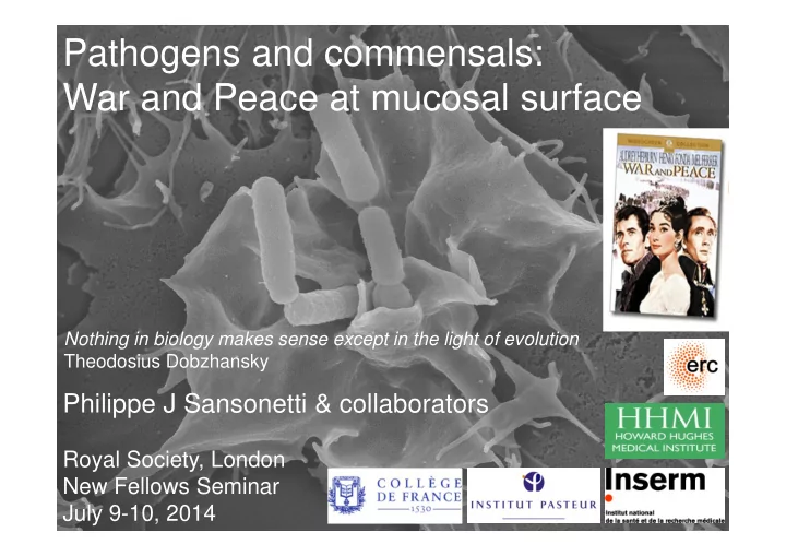
Pathogens and commensals: War and Peace at mucosal surface Nothing in biology makes sense except in the light of evolution Theodosius Dobzhansky Philippe J Sansonetti & collaborators Royal Society, London New Fellows Seminar July 9-10,
Innate immunity Physiological inflammation Surveillance/Tolerance Recognition network: PRRs:TLRs,NLRs, Rig1, MDA5…
Innate immunity Pathological inflammation Microbe & tissue destruction Amplification loop: TREM, HMGB1, Gal3, Sepsis, Septic shock Regulation Loss of control
Sansonetti, 2004, Nature Rev. Immunol. Sansonetti, 2006, Nat. Immunol. Sansonetti & Di Santo, 2007, Immunity Sansonetti & Medzhitov, 2009, Cell Sansonetti, 2010, Mucosal Immunology
Rupture of homeostasis = IBD,
Adaptive immunity Pathogens recognition, capture, completion of eradication process, protection
1012/ml in colon
O2, NO, ROS Antimicrobial peptides Lysozyme, proteases, lectins, phospholipases Transmigrating phagocytes sIgA Epithelium Mucus Cationic antimicrobial peptide hBD3 Sperandio et al.
Kim et al., PNAS, 2005 Arbibe et al., Nature Immunol., 2007 Sperandio et al., J. Exp. Med., 2008 Marteyn et al., Nature, 2010 Konradt et al., Cell Host Microbe, 2011 Puhar et al., Immunity, 2013
Symbionts/commensals: survive at distance or in particular niches, escape (regulate) innate defenses Pathogens: engage epithelium and subvert immune defenses
key immune signaling molecules
OCTN2 PepT1 QSM (hsl) MDP TLR NLR
Regulatory cascade
?
Regulatory genes
Mucus layer Regulatory cytokines, chemokines Antimicrobial molecules Epithelial apex Microbiota
Increase in ratio Treg/Th1, Th17 lymphocytes Increase in ratio Immature/mature DC and MΦ
Absence (limitation) of virulence factors PAMPs less agonist ? Sequestration, weak activity
Life in biofilms on mucus surface (at distance of epithelium) Controled diffusion and sampling of PAMPs and prokaryotic signalisation molecules
Symbionts
Pro-inflammatory cascade
TLR
Pro-inflammatory genes
PMN Activated DC & MΦ Th1 / Th17 Pro-inflammatory cytokines, chemokines PMN
DC
K+ K+ NLRs
Caspase-1 activation IL-1β Muropeptides Flagellin
1 – Mucinases Eradication
microbiota (niche occupancy) Adhesins / Invasins Secretory systems /effectors Hemolysins Massive engagement of PRRs Pathogenic properties sensed as exogenous danger signals 2 - Release of endogenous danger signals (DAMPs /small molecules) before initiation of proinflammatory transcriptional reprogramming. 3 – Subversion / dampening of innate (inflammatory) and adaptive immune responses
Epithelial cells mucus
Basolateral macropinocytosis (TTSS) Vacuole lysis (TTSS) Escape to autophagy Motility, cell to cell spread (TTSS) IcsA
? ?
M cell MΦ B Lympho MΦ pyroptosis TTSS/IpaB
DC «facilitated translocation» Follicle-associated epithelium PNN Nod1 PGN NF-κB JNK TTSS Pro-inflammatory genes IL-8, other cytokines chemokines
capacities Defensins and other bactericidal molecules CCL-20 B Lympho T Lympho
NOD LRRs CARD NOD LRRs CARD CARD
TLRs
NOD1 NOD2 IRAK TRAF6 RICK/Rip2
IkB IkB
P P
IkB IkB
P P
U U U U
MEKK3 IKK complex
IkB
PAMPs
Inflammatory programing based on sensing abnormal localisation of bacterial cell wall fragments and transcription of innate immunity genes
JNK Caspase-9
Girardin et coll., EMBO Reports, 2001 Girardin et coll.., Science, 2003 Girardin et coll., J.Biol.Chem., 2003
Muramyl-tri/tetrapeptide Muramyl-dipeptide
Inflammation
Pro-inflammatory genes Tri-Tetra-DAP/Nod1 MDP/Nod2
Pyd
HSP90
Pyd NBD
SGT1
LRR NLRP3 NBD NLRP3 Caspase NBD NBD NAIP5 NLRC4/IPAF Pro-caspase-1 NLRC4/IPAF inflammasome NLRP3 inflammasome Caspase-1 Pro-IL1β Pro-IL18 IL1β IL18
Apoptosis
?
P2X7 Pannexin-1 (K+) K+ Bacterial hemolysins IpaB IpaC Uric acid cristals TTSS translocator ATP Flagellin
S.typhimurium P.aeruginosa
Flagellin
L..pneumophila
?
S.flexneri
IL1β IL18
S.aureus L.monocytogenes A.hydrophyla
Nigericin Maitotoxin K+
Zychlinsky et al., 1992, Nature Zychlinsky et al., 1994, J Clin Invest Hilbi H et al., 1998, J Biol Chem
Macrophages
Rapid inflammatory programing based on release of a presynthetized pool of pro-inflammatory cytokines (Il-1β β β β, IL-18)
Inflammation
Inflammasome activation Differentiation of naive T cells to Th17 cells
HEMICHANNEL (Connexins)
x d x d x d x d x d x d x d x d x d x d x d x d x d x d x d x d x d x d x d x d x d x d x d x d x d x d x d x d x d x d x d x d x d x d x d x dx d x d x d x d x d x d x d x d x d x d x d x d x d x d x d x d x d x d x d x d x d x d x d x d x d x d
Tran Van Nhieu et al., 2003, Nat Cell Biol Puhar et al., 2013, Immunity
Rapid inflammatory programing based on release
ATP (DAMPs) = Inflammasome activation = Th17 lymphocytes differentiation
Inflammation
VirB
ipaA, ipaB, ipaC, ipaD, ipgB1, ipgD, icsB,
ipaH1/2, ipaH4, ipaH7, ipaH9.8
MxiE
INVASION MODULATION OF INNATE RESPONSES
IpaB, IpaC, IpaA, IpgB1,VirA, IpgD IpgD: phosphatidyl-inositol phosphatase, hydrolyses P in 4 in Pi(4,5)P2 (Niebuhr et al, 2002, Pendaries et al, 2006 EMBO J.). Anti-inflammatory +++ (Puhar et al., in review). OspG: kinase,binds/blocks ubiquitin transfer protein E2, protects I-kB from degradation. Anti-inflammatory +++ (Kim et al., 2005, PNAS). OspF: dephosphorylation of Erk1/2, epigenetic regulation of pro-inflammatory genes - i.e. IL-8. Regulates transmigration
Phosphothreonine lyase (Li et al., 2007, Science). IpaHs: (5 + 5 chromosomal copies): New family of Ubiquitin ligases (E3) (Rohde et al., 2007, Cell Host & Microbes) IpaH9.8 targets NEMO (Ashida et al., 2010, Nat.Cell Biol.)
OTHER PHENOTYPES
IcsB: inhibion or autophagy (Ogawa et al., 2005, Science) VirA: inhibition of microtubules, facilitates actin-based motility (Yoshida et al., 2006, Science)
virA
INHIBITION OF SECRETION
IpaB:(Mounier et al.,2012. Cell Host & Microbe)
HEMICHANNEL (Connexins)
x d x d x d x d x d x d x d x d x d x d x d x d x d x d x d x d
IpgD
Pi(4,5)P2 Pi(5)P IpgD
Pi(5)P
Niebuhr et al., Mol. Micro. 2000 Niebuhr et al., EMBO J. 2002 Pendaries et al., EMBO J 2006
Inflammation x d x dx d x d x d x d
ATP release in luminal fluid, rabbit ileal loop. 4h infection Puhar et al., 2013, Immunity
wt S. flexneri) IpgD-
Histopathology, HES Rabbit ileal loop 8 h infection
IpgD
cellules épithéliales mucus macropinocytose baso-latérale (TTSS) lyse vacuole (TTSS) motilité/passage cellule-cellule (TTSS) IcsA M cell MΦ Lympho B MΦ pyroptosis TTSS/IpaB
inflammatory apoptosis
DC «facilitated translocation» follicle-associated epithelium Nod1 PGN NF-κB JNK TTSS Osp(s) TTSS IL-8 CCL-20 PNN
Suppression of humoral defense mechanisms Suppression of cellular defense mechanisms
AMPs
Kim et al. 2005. PNAS Arbibe et al. 2007. Nat Immunol Sperandio et al.2008. J Exp Med Bergounioux et al. 2012. Cell Host Microbe Sperandio et al. 2013. Infect Immun Puhar et al. 2013. Immunity, ATP
ATP
Suppression of danger signaling PR
ATP
ATP
Laurence Arbibe Stéphane Girardin Benoît Marteyn F-Xavier Campbell-Valois Maria Mavris Joëlle Mounier Claude Parsot Armelle Phalipon Thierry Pédron Andrea Puhar John Rohde Brice Sperandio Pamela Schnupf REST OF THE WORLD Guy Tran Van Nhieu et al. (INSERM, Coll. de France)Laurent Combettes (INSERM, Orsay) Bernard Payrastre et al. (INSERM, Toulouse) Chris Tang (University of Oxford) Philippe Jay (INSERM, IGF, Montpellier) Françoise Poirier et al. (Institut Jacques Monod, Paris) Sylvie Robine et al. (Institut Curie, Paris) Dana Philpott Marie-C. Prévost Jean-Yves Coppée Christian Muchardt Jost Enninga Nathalie Sauvonnet