SLIDE 7 7
General Fluoroscopy Guidelines General Fluoroscopy Guidelines
- Physicians and Technologists should only radiate when necessary
Physicians and Technologists should only radiate when necessary and for as short a time as possible (i.e. Using pulsed fluorosc and for as short a time as possible (i.e. Using pulsed fluoroscopy)
- py)
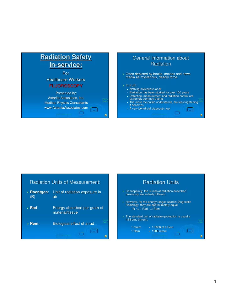
- Use automatic dose rate control.
Use automatic dose rate control.
- Collimate as much as possible.
Collimate as much as possible.
- Stand as far away as possible from the scatter radiation source,
Stand as far away as possible from the scatter radiation source, the the anatomy being imaged. anatomy being imaged.
Scatter on the X-
- ray tube side of the patient is much greater than on
ray tube side of the patient is much greater than on the II side of the patient. the II side of the patient.
- Wear aprons and other protective clothing as appropriate.
Wear aprons and other protective clothing as appropriate.
The x-
- ray tube to skin distance should be kept as large as possible
ray tube to skin distance should be kept as large as possible to reduce absorbed dose to the patient. This is accomplished by to reduce absorbed dose to the patient. This is accomplished by keeping the image intensifier as close to the patient as possibl keeping the image intensifier as close to the patient as possible. e.
- Only necessary personnel are to be in room during procedure.
Only necessary personnel are to be in room during procedure.
- Remove all supplementary objects from the primary beam (this
Remove all supplementary objects from the primary beam (this includes user hands). includes user hands).
Place the x-
- ray source under table for added user safety.
ray source under table for added user safety.
General Fluoroscopy Guidelines General Fluoroscopy Guidelines
- Physicians and Technologists should only radiate when necessary
Physicians and Technologists should only radiate when necessary and for as short a time as possible (i.e. Using pulsed fluorosc and for as short a time as possible (i.e. Using pulsed fluoroscopy)
- py)
- Use automatic dose rate control.
Use automatic dose rate control.
- Collimate as much as possible.
Collimate as much as possible.
- Stand as far away as possible from the scatter radiation source,
Stand as far away as possible from the scatter radiation source, the the anatomy being imaged. anatomy being imaged.
Scatter on the X-
- ray tube side of the patient is much greater than on
ray tube side of the patient is much greater than on the II side of the patient. the II side of the patient.
- Wear aprons and other protective clothing as appropriate.
Wear aprons and other protective clothing as appropriate.
The x-
- ray tube to skin distance should be kept as large as possible
ray tube to skin distance should be kept as large as possible to reduce absorbed dose to the patient. This is accomplished by to reduce absorbed dose to the patient. This is accomplished by keeping the image intensifier as close to the patient as possibl keeping the image intensifier as close to the patient as possible. e.
- Only necessary personnel are to be in room during procedure.
Only necessary personnel are to be in room during procedure.
- Remove all supplementary objects from the primary beam (this
Remove all supplementary objects from the primary beam (this includes user hands). includes user hands).
Place the x-
- ray source under table for added user safety.
ray source under table for added user safety.