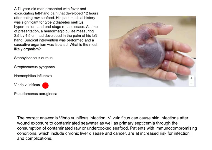
SLIDE 1 A 71-year-old man presented with fever and excruciating left-hand pain that developed 12 hours after eating raw seafood. His past medical history was significant for type 2 diabetes mellitus, hypertension, and end-stage renal disease. At time
- f presentation, a hemorrhagic bullae measuring
3.5 by 4.5 cm had developed in the palm of his left
- hand. Surgical intervention was performed and a
causative organism was isolated. What is the most likely organism? Staphylococcus aureus Streptococcus pyogenes Haemophilus influenza Vibrio vulnificus Pseudomonas aeruginosa
The correct answer is Vibrio vulnificus infection. V. vulnificus can cause skin infections after wound exposure to contaminated seawater as well as primary septicemia through the consumption of contaminated raw or undercooked seafood. Patients with immunocompromising conditions, which include chronic liver disease and cancer, are at increased risk for infection and complications.
SLIDE 2 Vibrio vulnificus ist eine Art gram-negativer, gekrümmter Stäbchen-Bakterien der Gattung Vibrio. Sie kommen vor in marinen Umgebungen wie Flussmündungen, Brackwasser-Tümpeln oder Küstengebieten. V. vulnificus ist eng verwandt mit V. cholerae, dem Auslöser der Cholera. Eine Infektion mit V. vulnificus führt zu sich rasch ausdehnender Cellulitis oder Sepsis. Vibrio vulnificus verursacht eine Infektion, die häufig nach dem Verzehr von Meeresfrüchten, insbesondere Austern, auftritt; die Bakterien können auch durch
- ffene Wunden (dazu zählen auch frische, d. h. noch
nicht vollständig verheilte Tätowierungen) in den Körper eindringen beim Schwimmen oder Waten in verseuchten Gewässern, oder über Stichwunden durch Dornen von Fischen wie den Tilapia. Zu den Symptomen gehören Erbrechen, Diarrhö, Leibschmerzen und eine Blasen werfende Dermatitis, die manchmal fälschlicherweise für Pemphigus vulgaris
- der Pemphigus gehalten wird.
Bei Menschen mit geschwächtem Immunsystem wie chronischer Leberkrankheit kann sich ein mit Vibrio- Bakterien infizierter Schnitt rasch verschlimmern und auf den Blutkreislauf übergreifen. Schwere Symptome können auftreten, sogar Tod.
SLIDE 3 Clopidogrel and Aspirin in Acute Ischemic Stroke and High-Risk TIA
Combination antiplatelet therapy with clopidogrel and aspirin may reduce the rate of recurrent stroke during the first 3 months after a minor ischemic stroke or transient ischemic attack (TIA). A trial of combination antiplatelet therapy in a Chinese population has shown a reduction in the risk of recurrent stroke. We tested this combination in an international
- population. In a randomized trial, we assigned
patients with minor ischemic stroke or high-risk TIA to receive either clopidogrel at a loading dose of 600 mg on day 1, followed by 75 mg per day, plus aspirin (at a dose of 50 to 325 mg per day) or the same range of doses of aspirin
- alone. The dose of aspirin in each group was
selected by the site investigator. The primary efficacy outcome in a time-to-event analysis was the risk of a composite of major ischemic events, which was defined as ischemic stroke, myocardial infarction, or death from an ischemic vascular event, at 90 days. Patients who were at least 18 years of age were enrolled if they could undergo randomization within 12 hours after having an acute ischemic stroke with a score of 3 or less on the National Institutes of Health Stroke Scale (NIHSS) (scores range from 0 to 42, with higher scores indicating greater stroke severity) or a high-risk TIA with a score of 4 or more on the ABCD2 scale
SLIDE 4
Shown are the percentages of patients with the primary efficacy outcome (a composite of ischemic stroke, myocardial infarction, or death from ischemic vascular causes) (Panel A) and the primary safety outcome of major hemorrhage (Panel B). Inset graphs show the same data on an expanded y axis.
SLIDE 5
SLIDE 6
SLIDE 7 In this international, multicenter, randomized trial, we found that patients with minor ischemic stroke or high-risk TIA who received a combination of clopidogrel and aspirin had a lower risk of major ischemic events but a higher risk of major and minor hemorrhage than did those receiving aspirin alone. Ischemic stroke accounted for most
- f the composite events of the primary efficacy outcome, and the effect of dual
antiplatelet treatment was attributable to a reduction in the rate of these strokes. It is not possible to make direct comparisons between clinical and safety outcomes because disability due to each of the outcomes cannot be ascertained, but we estimate that for every 1000 patients who are treated with clopidogrel plus aspirin during a period of 90 days, such treatment would prevent approximately 15 ischemic events and would cause 5 major hemorrhages. The aspirin dose varied in the two treatment groups, which reflected the clinical practices of local investigators; however, in a potentially underpowered analysis, no difference in treatment effect was shown across aspirin doses. In conclusion, in patients from diverse countries with minor ischemic stroke or high-risk TIA, those who received a combination of clopidogrel and aspirin had a lower risk of a composite of ischemic stroke, myocardial infarction, or death from ischemic vascular causes but had a higher risk of major hemorrhage than patients who received aspirin alone during the 90-day trial period.
SLIDE 8 Die Sichelzellanämie ist eine Erbkrankheit, die zu den hämolytischen Anämien bzw. Hämoglobinopathien gehört. Sie wird durch einen genetischen Defekt (Punktmutation) ausgelöst, der zur Bildung von irregulärem Hämoglobin, dem so genannten Sichelzellhämoglobin (Hämoglobin S, HbS) führt. Die Sichelzellanämie folgt einem autosomal-rezessiven
- Erbgang. Es besteht eine Punktmutation der ß-Globinkette,
wobei an Position 6 hydrophiles Glutamat gegen hydrophobes Valin substituiert wurde. Im desoxygenierten Zustand des HbS bilden sich Aggregate und es kommt zur Hämolyse. Es erkranken nur homozygote Merkmalsträger, bei denen das mütterliche und väterliche Gen verändert ist. Heterozygote Merkmalsträger sind weitgehend symptomfrei, da beide Allele kodominant sind, d.h. die Merkmalsträger produzieren sowohl normales als auch defektes Hämoglobin. Allerdings wird auch bei ihnen unter starkem Sauerstoffmangel, wie zum Beispiel während einer Narkose, die Sichelform der Erythrozyten ausgebildet, so dass es zu einer Beeinträchtigung der Organdurchblutung kommen kann. Das akute Krankheitsgeschehen der Sichelzellanämie findet während einer sogenannten Sichelzellkrise statt. Auslöser einer Sichelzellkrise sind beispielsweise: Die Mikrozirkulation wird eingeschränkt und Organschädigungen resultieren. Nach längerer Krankheitsdauer können sich bedrohliche Komplikationen entwickeln, darunter Infarkte von Organen (z.B. Milz, Lunge). Zusätzlich kann sich ein akutes Thorax- Syndrom (ATS), ein Hand-Fuß-Syndrom (v.a. im Kindesalter)
- der ein Priapismus klinisch manifestieren.
SLIDE 9 Hydroxycarbamid (oder hydroxyurea) ist ein zytostatisch wirkender Arzneistoff, dessen Einsatzschwerpunkt die Therapie der chronisch lymphatischen Leukämie (CLL) darstellt. Seltener wird der Wirkstoff auch gegen die Sichelzellenanämie oder die Thalassämia major verwendet. Hydroxycarbamid wirkt hemmend auf die Mitose und die DNA-Synthese. Hydroxycarbamid hemmt zunächst einmal die Synthese von DNA, was eine Zellteilung unmöglich macht. Der genaue Wirkungsmechanismus ist noch nicht gänzlich geklärt. Als wahrscheinlich gilt, dass das Zytostatikum das Enzym Ribonukleotidreduktase hemmt. Dieses katalysiert die Umwandlung von Ribose in
- Desoxyribose. Letztere ist ein wichtiger Baustein der
- DNA. Steht nicht genug Desoxyribose zur Verfügung,
ist DNA-Synthese blockiert. Gleiches gilt folglich für die Zellteilung. Hydroxycarbamid verhindert weiterhin eine Integration von Thymin-Nukleotiden in den DNA-Strang. Fetales Hämoglobin stoppt das Entstehen dieser Polymere im Sichelhämoglobin innerhalb der roten Blutkörperchen. Das Medikament Hydroxyurea wird verwendet, um fetales Hämoglobin zu erhöhen und dadurch die Auswirkungen der Erkrankung zu reduzieren. Dies ist eine Aktualisierung eines zuvor veröffentlichten Cochrane Review.
SLIDE 10 A Phase 3 Trial of l-Glutamine in Sickle Cell Disease
Oxidative stress contributes to the complex pathophysiology of sickle cell disease. Oral therapy with pharmaceutical-grade l-glutamine (USAN, glutamine) has been shown to increase the proportion of the reduced form of nicotinamide adenine dinucleotides in sickle cell erythrocytes, which probably reduces
- xidative stress and could result in fewer episodes of
sickle cell–related pain. In a multicenter, randomized, placebo-controlled, double-blind, phase 3 trial, we tested the efficacy of pharmaceutical-grade l-glutamine (0.3 g per kilogram of body weight per dose) administered twice daily by mouth, as compared with placebo, in reducing the incidence of pain crises among patients with sickle cell anemia or sickle β0-thalassemia and a history of two or more pain crises during the previous year. Patients who were receiving hydroxyurea at a dose that had been stable for at least 3 months before screening continued that therapy through the 48- week treatment period. Oxidative stress contributes to the complex pathophysiology of sickle cell disease. Nicotinamide adenine dinucleotide (NAD+) is a ubiquitous oxidation– reduction (redox) cofactor in red cells. NAD+ and its reduced form, NADH, play major roles in maintaining redox balance. Sickle red cells have a lower redox ratio ([NADH]:[NAD++NADH]) than normal red cells.
SLIDE 11 Recurrent Events of Sickle Cell–Related Pain Crisis over Time, According to Trial Group. An analysis of sickle cell– related pain crisis over time yielded an intensity rate ratio (i.e., the ratio of the recurrent event rates in each trial group)
- f 0.75 (95% CI, 0.62 to 0.90,
according to the Andersen–Gill model; and 95% CI, 0.55 to 1.01, according to the Lin– Wei–Yang–Ying modification of the Andersen–Gill model), which indicates that the cumulative number of painful crises was 25% lower in the l- glutamine group than in the placebo group over the entire 48-week treatment period.
SLIDE 12 Time to Sickle Cell–Related Pain Crisis. Panel A shows the Kaplan–Meier curves for time to the first sickle cell–related pain
- crisis. The median time to the first pain
crisis was 84 days (95% CI, 62 to 109) in the l-glutamine group, as compared with 54 days (95% CI, 31 to 73) in the placebo group (hazard ratio, 0.69; 95% CI, 0.52 to 0.93; P=0.02). Panel B shows the Kaplan–Meier curves for time to the second sickle cell–related pain crisis. The median time to the second pain crisis was 212 days (95% CI, 153 to 250) in the l- glutamine group, as compared with 133 days (95% CI, 115 to 179) in the placebo group (hazard ratio, 0.68; 95% CI, 0.49 to 0.96; P=0.03).
SLIDE 13
SLIDE 14
The rate of adverse events was higher in the placebo group than in the l- glutamine group (100% vs. 98.0%), as was the rate of serious adverse events (87.1% vs. 78.2%). Adverse events with a higher incidence in the l- glutamine group than in the placebo group and with at least a 5% incidence in the l-glutamine group are listed.
SLIDE 15
In the current trial, in which the majority of patients received concomitant hydroxyurea, the number of pain crises per patient was significantly lower in the l-glutamine group than in the placebo group and differed between trial groups by a median of one event over 48 weeks. The time to the first pain crisis began to diverge within 2 weeks after the start of the treatment period, with sustained separation of curves over the duration of the trial. The analysis of recurrent pain crises over time reinforced the observation that over the entire trial period, the median number of pain crises was 25% lower with l-glutamine than with placebo. The exact mechanisms by which l-glutamine reduces the frequency of pain crises have not been fully elucidated. The 33% between-group difference in the median number of hospitalizations is notable because hospitalization can be very costly. The mean number of ED visits was lower in the l-glutamine group than in the placebo group, but the difference was not significant. In 2017, the U.S. Food and Drug Administration approved l-glutamine (USAN, glutamine) for the prevention of acute vaso-occlusive pain events in persons with sickle cell disease who are older than 5 years of age — only the first drug to be approved for this indication in the 20 years since the approval of hydroxyurea. The price of l-glutamine is much higher than that of hydroxyurea; 1 year of treatment with Endari (Emmaus Medical) for an average adult is estimated at $40,515, as compared with approximately $1700 for hydroxyurea. Whether the cost will be a hindrance to its use has yet to be determined. This agent certainly has been slow to enter the market because prescribing l- glutamine for patients requires many steps, which may dissuade busy practitioners from actively prescribing it. Because l-glutamine has a putatively different mechanism (or mechanisms) of action and toxicity profile than hydroxyurea, concomitant use is possible and most likely advantageous. Therefore, caution may be warranted in prescribing l-glutamine to patients with sickle cell disease who have clinically significant renal and hepatic dysfunction. Of note, in the two randomized trials of l-glutamine involving patients with sickle cell disease, three deaths occurred in the l-glutamine groups, as compared with none in the placebo groups.
SLIDE 16 Procalcitonin ist das Prohormon des Calcitonins. Unter physiologischen Bedingungen wird Procalcitonin in den C-Zellen der Schilddrüse als Vorläuferprotein gebildet und proteolytisch zum funktionsfähigen Hormon
- prozessiert. Bei einem generalisiert-entzündlichen
Geschehen wird Procalcitonin aus bisher nicht verstandenen Gründen vermehrt gebildet. Anders als unter physiologischen Bedingungen erfolgt die Synthese dann aber vermutlich in der Leber. Freisetzungsreiz sind hierbei u.a. bakterielle Endotoxine, z.B. das Lipopolysaccharid. Procalcitonin erlangt dadurch eine hohe Spezifität für bakterielle Infektionen, insbesondere bei Vorliegen einer Sepsis oder systemisch verlaufenden
- Erkrankungen. In diesen Fällen kann es auf Werte von
10-1000 μg/l ansteigen. Es kann jedoch auch zu einem Anstieg des Procalcitonin-Wertes bei Pilzinfektionen und protozoenbedingten Infektionen kommen. Bei Virus- oder Autoimmunerkrankungen hingegen steigt es nicht oder nur marginal an. Durch dieses Verhalten leistet das Procalcitonin wertvolle Dienste hinsichtlich der Diskriminierung von bakteriellen und viralen Entzündungen. Serumspiegel < 0,5 μg/ll
SLIDE 17
Procalcitonin-Guided Use of Antibiotics for Lower Respiratory Tract Infection
The effect of procalcitonin-guided use of antibiotics on treatment for suspected lower respiratory tract infection is unclear. In 14 U.S. hospitals with high adherence to quality measures for the treatment of pneumonia, we provided guidance for clinicians about national clinical practice recommendations for the treatment of lower respiratory tract infections and the interpretation of procalcitonin assays. We then randomly assigned patients who presented to the emergency department with a suspected lower respiratory tract infection and for whom the treating physician was uncertain whether antibiotic therapy was indicated to one of two groups: the procalcitonin group, in which the treating clinicians were provided with real-time initial (and serial, if the patient was hospitalized) procalcitonin assay results and an antibiotic use guideline with graded recommendations based on four tiers of procalcitonin levels, or the usual-care group. We hypothesized that within 30 days after enrollment the total antibiotic-days would be lower — and the percentage of patients with adverse outcomes would not be more than 4.5 percentage points higher — in the procalcitonin group than in the usual-care group.
SLIDE 18
Violin plots for the primary outcome of antibiotic-days by day 30 are shown. The width of the colored shape indicates the probability density of patients with a given result. The gray notched box plots represent the median (yellow horizontal line), 95% confidence interval of the median (notch), interquartile range (25th to 75th percentile) (box), and the upper 1.5 times the interquartile range (solid vertical line).
SLIDE 19
Antibiotic Exposure. In the intention-to-treat analysis, there was no significant difference in antibiotic exposure during the first 30 days between the procalcitonin group and the usual-care group (mean antibiotic-days, 4.2 and 4.3 days, respectively; difference, −0.05 day; 95% confidence interval [CI], −0.6 to 0.5; P=0.87). The results were similar in the per-protocol analysis (difference, −0.1 day; 95% CI, −0.7 to 0.6), per- guideline analysis (−0.1 day; 95% CI, −1.0 to 0.8), complete-case analysis (−0.1 day; 95% CI, −0.7 to 0.5), and missing-not-at-random analysis (−0.1 day; 95% CI, −0.7 to 0.5). There was no significant difference in antibiotic-days by day 30 in any prespecified subgroup analysis.
SLIDE 20
Antibiotic Exposure over Time. Day 1 is from the time of enrollment to midnight. Day 2 and beyond are from midnight to midnight. Serial procalcitonin levels were obtained from hospitalized patients through day 7. Data on post-discharge antibiotic exposure were derived from the intention-to-treat analysis of the primary outcome.
SLIDE 21 In this multicenter trial, the use of a procalcitonin-guided antibiotic prescription guideline did not result in less exposure to antibiotics than did usual care among patients presenting to the emergency department with suspected lower respiratory tract infection. There are several possible explanations for this finding. In the usual-care group, even when clinicians did not know the procalcitonin assay result, they prescribed antibiotics less frequently to patients in the lower procalcitonin-level tiers than to those in the higher tiers. Patients with lower procalcitonin levels also had fewer clinical features of infection, and in that context, procalcitonin probably provided a modest amount of additional information to guide decisions. There was some suggestion of heterogeneity of the effect of the intervention, such as lower antibiotic prescription rates for patients with acute bronchitis than for those with other final diagnoses and a possible interaction between treatment effect and procalcitonin tier. However, these secondary analyses were exploratory, and the differences were largely nonsignificant. We did not directly address whether antibiotics can be safely withheld on the basis of a low procalcitonin level alone but rather tested the effect of a deployment strategy to promote the recommended use of the assay in clinical practice (in a patient population in which the likelihood
- f antimicrobial use was intermediate). In our strategy, procalcitonin assay results were provided
to the clinical team before decision making in most but not all instances. A lack of knowledge about the prescribing practices used by individual physicians limits the insights we can make. The potential effect of emerging technology that may improve the rapid identification of infectious agents — technology that was largely unavailable during the course of this trial — is
- unclear. Finally, we did not achieve follow-up for all the patients in our trial, but our results were
robust to complete-case and missing-not-at-random sensitivity analyses.
SLIDE 22 Physiologisch ist die Koronare Flussreserve (CFR von coronary flow reserve) der Quotient des maximalen Blut- Volumenstroms in den Koronararterien unter Belastung und des Blut-Volumenstroms in den Koronararterien in
- Ruhe. Es ist also der maximale zusätzliche Volumenstrom, der den Herzmuskel versorgen kann, wenn es belastet
wird und einen erhöhten Bedarf an Blutversorgung hat. Sie wird üblicherweise als Quotient von maximalem Belastungs- und Ruhe-Volumenstrom angegeben. Beim Gesunden kann die CFR den Wert 6 oder höher betragen; als normal werden Werte oberhalb von 3,5
- angesehen. Die CFR ist unter anderem erniedrigt bei Stenosen in den Koronararterien, bei Bluthochdruck, beim
Kardialen Syndrom X und bei der Hypertrophen Kardiomyopathie. Goldstandard zur Bestimmung der Koronaren Flussreserve ist die Untersuchung mit einem intrakoronaren Doppler-Draht im Rahmen einer Herzkatheteruntersuchung. Des Weiteren werden PET sowie die transthorakale Echokardiografie bei der LAD eingesetzt. Allen Methoden gemeinsam ist, dass zunächst eine Untersuchung in Ruhe durchgeführt wird und dann unter medikamentöser Belastung wiederholt wird, zumeist mit Adenosin.
SLIDE 23 Five-Year Outcomes with PCI Guided by Fractional Flow Reserve
We hypothesized that fractional flow reserve (FFR)–guided percutaneous coronary intervention (PCI) would be superior to medical therapy as initial treatment in patients with stable coronary artery disease. Among 1220 patients with angiographically significant stenoses, those in whom at least
- ne stenosis was hemodynamically
significant (FFR, ≤0.80) were randomly assigned to FFR-guided PCI plus medical therapy or to medical therapy alone. Patients in whom all stenoses had an FFR of more than 0.80 received medical therapy and were entered into a
- registry. The primary end point was
a composite of death, myocardial infarction, or urgent revascularization.
SLIDE 24
SLIDE 25
Kaplan–Meier Curves for the Primary End Point. Shown is the cumulative incidence of the primary end point (a composite of death from any cause, myocardial infarction, or urgent revascularization) in the two groups in the trial. A hazard ratio below 1.00 denotes a lower incidence of the primary end point in the group that underwent fractional flow reserve– guided percutaneous coronary intervention (PCI) than in the medical-therapy group.
SLIDE 26 Kaplan–Meier Curves for Death from Any Cause, Myocardial Infarction, and Urgent Revascularization. Hazard ratios below 1.00 denote a lower incidence of events in the PCI group than in the medical-therapy group. The 95% confidence intervals for secondary end points were not adjusted for multiple testing, and any inferences drawn from the intervals as reported may not be
- reproducible. Insets show the same data
- n an enlarged y axis.
SLIDE 27 Angina Class in Patients in the Trial Groups and Registry Cohort over Time. Shown are the numbers of patients in the two trial groups and the registry cohort who had angina of class II to IV on the Canadian Cardiovascular Society (CCS) scale (which ranges from I to IV, with higher classes indicating greater limitations on physical activity
- wing to angina) at various time points. The 95% confidence intervals for secondary end points were not adjusted for multiple testing,
and any inferences drawn from the intervals as reported may not be reproducible.
SLIDE 28 This 5-year follow-up of the FAME 2 trial showed that, among patients with stable angina, FFR- guided PCI led to a significantly lower rate of the prespecified primary composite end point of death, myocardial infarction, or urgent revascularization than medical therapy alone. This difference was driven by a significantly lower rate of urgent revascularization in the PCI group than in the medical-therapy group. Patients in whom all coronary stenoses were hemodynamically nonsignificant had an event rate with medical therapy alone that did not differ significantly from the rate among patients with hemodynamically significant stenoses who underwent FFR-guided PCI. There was no evidence of convergence of event rates between groups in the long term. Patients who had originally been assigned to undergo FFR-guided PCI reported significantly less angina up to 3 years after randomization than did patients who had been assigned to receive medical therapy
- alone. However, this difference was no longer significant at 5 years, by which time 51% of the
patients who had been initially assigned to medical therapy alone had undergone revascularization. Some limitations must be taken into account. First, enrollment was stopped prematurely by the data and safety monitoring board because of a large excess of primary end-point events in the medical-therapy group. The early termination of clinical trials has been shown to exaggerate treatment effects. In conclusion, in patients with stable coronary artery disease, an initial FFR-guided PCI strategy resulted in a sustained clinical benefit, as compared with medical therapy alone, with regard to the composite primary end point of death, myocardial infarction, or urgent revascularization at 5 years. Patients without hemodynamically significant stenoses had a favorable long-term outcome with medical therapy alone.
SLIDE 29
A 36-year-old man presented to the emergency department with a 2-week history of fever, headache, drowsiness, and photophobia. He was previously healthy and was sexually active with men. The physical examination was notable for a temperature of 38.3°C and neck stiffness. Computed tomography of the head was normal. The opening pressure on lumbar puncture was 29 cm of water (reference range, <20 cm). The cerebrospinal fluid (CSF) cell count was 340 cells per microliter (reference range, 0 to 10), with 90% mononuclear cells, which were predominantly lymphocytes. The glucose level was 46 mg per deciliter (2.6 mmol per liter; reference range, 40 to 70 mg per deciliter [2.2 to 3.8 mmol per liter]), and the protein level was 0.80 g per liter (reference range, 0.15 to 0.45). Gram’s stain (Panel A) and India ink stain (Panel B) revealed abundant encapsulated, round yeasts, with some budding forms. The cryptococcal antigen titer was 1:128, and the CSF culture grew Cryptococcus neoformans. No other pathogen was detected. A test for the human immunodeficiency virus antibody was positive; the viral load was 300,000 copies per milliliter, and the CD4+ count was 7 cells per microliter (reference range, 500 to 1450). Induction therapy with liposomal amphotericin B and flucytosine was started, and resolution of symptoms and negative results on CSF culture were noted after 2 weeks of treatment. Consolidation therapy with fluconazole was started, and antiretroviral therapy was later prescribed.
SLIDE 30 Psychotic Disorders
The term “psychosis,” which is derived from the Greek word for abnormal condition of the mind, has been used in many different ways in clinical
- medicine. Before 1980, the term “psychotic” was
applied generically to persons whose mental functioning was sufficiently impaired to interfere with their capacity to meet the ordinary demands of life. Starting in 1980 with the publication of the third edition of the Diagnostic and Statistical Manual of Mental Disorders (DSM-III), the term indicated gross impairment in reality testing — that is, disruption of the ability to distinguish between the internal experience of the mind and the external reality of the environment. To achieve greater diagnostic precision, DSM-IV, published in 1994, defined psychosis more specifically to apply to mental disorders characterized by symptoms such as fixed false beliefs (delusions, such as the belief that one is being poisoned by neighbors who are piping gas through the walls), hallucinations, disorganized thoughts (illogical and incoherent speech, neologisms, and made-up words), clang associations (rhymed words), word salad (nonsensical sentences), echolalia (repetition
- f spoken words), and abnormal motor behavior
(bizarre postures, stereotypy, and waxy flexibility).
SLIDE 31
Natural History of Schizophrenia and the Rationale for Preventing Chronic Disease. Shown are the stages of illness in schizophrenia, the prototypical idiopathic psychotic disorder. Detection and treatment in the early stages of illness, ideally close to the onset of the first episode of psychosis, shorten the duration of psychotic episodes, reduce recurrences, and limit the progressive decline in functioning (deterioration) that occurs in the syndromal stage and leads to the chronic effects of the disease. The syndromal stage begins with the first episode of psychosis and continues through the progressive stage.
SLIDE 32 A sagittal view of the brain through the midline depicts the hippocampus, midbrain, corpus striatum, and prefrontal cortex, all regions that are implicated in psychotic symptoms and disorders. The affected neurotransmitters include dopamine (blue arrows), glutamate (purple arrows), and γ-aminobutyric acid (GABA) (green arrows). Idiopathic psychoses (e.g., schizophrenia and mood disorders with psychotic symptoms) are believed to arise from overactivity of neurons that release glutamate onto cells located in and projecting from the CA1 region of the hippocampus. Deficits in hippocampal GABAergic interneurons and hypofunctioning N- methyl-D-aspartate glutamate receptors (NMDARs) (the red X denotes hypofunction) are the main molecules that are thought to be responsible for these disturbances. Shown in the hippocampus is a glutamate-expressing pyramidal cell and a GABA
- interneuron. Despite the increase in synaptic glutamate, there is underactivation of the interneuron. Also shown is the interneuron
axon forming a synapse with the apical dendrite of the pyramidal cell. Because of understimulation, the interneuron releases less GABA, which in turn disinhibits the pyramidal cell and causes it to release more glutamate from hippocampal projections to the midbrain (ventral tegmental area [VTA]) and the corpus striatum (nucleus accumbens). Hippocampal overactivity augments dopamine release in the striatum either directly (at the level of the nucleus accumbens) or by stimulation of midbrain dopamine neurons, which project to the nucleus accumbens and the prefrontal cortex. Dopamine neurons in the midbrain further promulgate the dysregulation of dopamine and glutamate through a projection back to the hippocampus. Psychotic symptoms can be induced in nonidiopathic disorders that affect these pathways at various locations. For example, in autoimmune or toxic psychoses (e.g., psychoses due to PCP), the exogenous drug or antibody acts as an NMDAR antagonist, thus mimicking a constitutionally hypofunctioning NMDAR. In Alzheimer’s disease, the cholinergic system is compromised and cholinergic inhibitory inputs to glutamateric cells in the hippocampus and dopaminergic cells in the midbrain are diminished. D2R denotes D2 receptor.
SLIDE 33
Approximately 20 antipsychotic medications are currently marketed in the United States, all of which work largely by blocking or mitigating the activity of dopamine at D2 receptors. These medications are effective in treating psychotic symptoms in patients with various disorders, although their effectiveness depends on their safety profile, which varies with the drug’s pharmacology and the nature of the underlying cause of the condition (e.g., dementia vs. drug- induced psychosis). The older — typical, or first-generation — medications have a propensity to cause extrapyramidal neurologic side effects, whereas the atypical, or second-generation, drugs are more likely to induce weight gain and disturbances in glucose and lipid metabolism. An exception is clozapine, which produces few extrapyramidal effects and has therapeutic efficacy in patients with a partial response or no response to other antipsychotic agents.32 However, clozapine can be associated with serious side effects, including seizures (in approximately 4% of patients), myocarditis (in 1%), and agranulocytosis (in 0.8%), and is therefore indicated mainly for the treatment of refractory psychotic symptoms. Enthusiasm for early intervention in and prevention of idiopathic psychotic disorders has led investigators and the National Institute of Mental Health to determine how to extend this approach to patients in the prodromal stage of illness in order to prevent the onset of a more disturbing psychotic disorder. Before this approach can be applied, better diagnostic methods must be developed, because the current criteria for identifying persons with attenuated psychotic symptoms who are believed to have a high clinical risk of conversion to a syndromal form of psychosis have a false positive rate that is higher than 50%. Consequently, a diagnostic test is needed that can identify psychotic episodes that will progress to a syndromal psychosis, as well as episodes that are stable or transient and those that can be attenuated. Treatments so far have been shown to be effective for alleviation of symptoms but not for prevention of conversion to syndromal psychosis.
SLIDE 34 A 47-year-old man presented to the emergency department with a 6-month history of worsening exertional
- dyspnea. He had recently emigrated from Honduras and had a history of cardiac surgery as a child, but the
initial diagnosis and specific procedure that had been performed were not known. The physical examination revealed a right parasternal heave, a loud S1, a midpeaking systolic ejection murmur (grade 2/6) at the upper sternal border, and a low-pitched diastolic rumble at the apex. An electrocardiogram showed atrial flutter with variable atrioventricular conduction. Transthoracic echocardiography revealed moderate aortic stenosis (calculated valve area, 1.3 cm2) and very severe mitral stenosis (calculated valve area, 0.5 cm2) (see video and Panel A; LA denotes left atrium, LV left ventricle, and MV mitral valve). The left atrium was enlarged, with an indexed atrial volume of 364 ml per square meter (normal value, <35). Color-flow Doppler images (Panel B, upper half) and a continuous-wave Doppler flow pattern (Panel B, lower half) showed the stenotic mitral valve during diastole. The continuous-wave Doppler flow pattern depicts flow velocity (vertical axis) and time (horizontal axis). The slow decline in transmitral flow velocity reflects the very slow decrease in left atrial pressure and slow increase in left ventricular diastolic pressure that occur in mitral stenosis. The mean transmitral gradient was 14 mm Hg (normal value, <3). The patient underwent replacement of the mitral and aortic valves with mechanical prostheses. At a 6-month follow-up visit, he had only slight limitations on physical activity (New York Heart Association class II).
SLIDE 35 A 64-year-old man was admitted to this hospital because of progressive leg weakness, recurrent falls, and anemia. Four months before this admission, the patient fell in his garage and attributed the fall to tripping over an object on the ground. He did not hit his head, lose consciousness, or need assistance to stand or walk after the fall. He was evaluated by his primary care provider. On evaluation, he reported 3 months of increased alcohol consumption (up to 6 or 7 glasses of wine each night) and several months of voluntarily restricted food intake to achieve weight loss. On physical examination, the vital signs were normal. The height was 166.6 cm, and the weight was 128.4 kg (6 months earlier, the weight had been 140.6 kg); the body-mass index (the weight in kilograms divided by the square
- f the height in meters) was 46. On evaluation in the clinic, the
patient reported mild pain above the left eye but no headache, neck pain, back pain, urinary retention, fecal incontinence, fever, chills, morning stiffness, muscle pain, muscle swelling, dark stools, melena, or hematemesis. He had a history of atrial fibrillation, hypertension, gout, osteoarthritis of the knees, gastroesophageal reflux disease, and Barrett’s esophagus, which had been diagnosed by means of biopsy 7 years before this admission. Roux-en-Y gastric bypass had been performed 6 years before this admission. Medications were allopurinol, amlodipine, furosemide, indomethacin, losartan, metoprolol,
- meprazole, rivaroxaban, bupropion, cyanocobalamin,
ergocalciferol, and a thiamine supplement. The patient was divorced, lived alone, and worked as a store manager. He did not smoke tobacco. His father and brother had both died of esophageal cancer.
SLIDE 36 The first and second heart sounds were normal, without murmurs. The breath sounds were normal bilaterally, without wheezing or
- rhonchi. Bowelsounds were present, and the
abdomen was soft, nondistended, and nontender on palpation. The edge of the liver was not palpable, and the spleen was not
- enlarged. Motor strength was 3 out of 5
bilaterally on hip flexion and 4 out of 5 bilaterally on hip extension; the patient could not rise from a seated position, even when he used his arms for assistance. The remainder of the motor examination was normal. There was no muscle atrophy, swelling, or tenderness on
- palpation. Perception of pinprick was
diminished in the legs from the toes to above the knees. Perception of light touch was diminished on the plantar surface of the feet. Proprioception was decreased in the big toes. Reflexes were normal, as were the results of finger–nose–finger testing. An evaluation for the Babinski sign and a Romberg test were not
- performed. Hair was thin and fragile on the
arms and absent on the legs. The stool was brown and negative for occult blood. The remainder of the physical examination was
- normal. The patient was referred to the
emergency department of this hospital for further evaluation.
SLIDE 37 The patient was admitted to the hospital, and a high-dose thiamine infusion, a folate supplement, and a multivitamin were administered, in addition to his usual medications. One hour after admission, the blood pressure was 89/47 mm Hg and the heart rate was 90 beats per minute while the patient was sitting; after he stood up, the blood pressure was 75/40 mm Hg and the heart rate was 100 beats per minute. Amlodipine, furosemide, losartan, and metoprolol were discontinued, and fluids were administered intravenously. Two days later, the blood pressure was 124/72 and the heart rate was 91 beats per minute while the patient was sitting; after he stood up, the blood pressure was 80/50 mm Hg and the heart rate was 89 beats per minute. He could rise from a seated position with minimal use of his arms. An area of bruising (10 cm in diameter) developed on the right forearm, where a peripheral intravenous catheter had been placed. Falls in Older Patients This patient attributed his fall to tripping over an object in his garage. After a fall, many patients develop a narrative or explanation to normalize the event. Falling is always a surprise to a person with a lifetime of experience remaining upright, despite tripping or walking on slippery surfaces. Maintaining balance after tripping is dependent on strength in the hip flexor and gluteal muscles, and such strength decreases gradually with aging. When patients describe a fall as “just tripping,” I ask whether they think they would have fallen if the same event had occurred 10 or 20 years earlier. Orthostatic Hypotension Could this patient’s recurrent falls have been due to orthostatic (postural) hypotension? An assessment for
- rthostatic hypotension would be useful, since he was receiving antihypertensives and diuretics, and medication
side effects such as orthostatic hypotension are another common cause of falls in older adults. On admission to the hospital, he was given his usual medications, as well as a standard multivitamin, a folate supplement, and a thiamine infusion. Rapid intravenous infusion of thiamine can cause hypotension, but such hypotension is
- transient. However, although this patient had potential age-related risk factors for falling, his alcohol use was the
most worrisome risk factor. Falls in Patients with Alcohol Use If I had seen this patient in the clinic, I would have strongly suspected that his first fall had been related to alcohol intoxication, since he had reported drinking up to 6 or 7 glasses of wine daily during the 3 months before the
- evaluation. Alcohol-use disorder can occur after Roux-en-Y gastric bypass in patients who have no history of
excessive drinking.
SLIDE 38 Falls in Patients with Nutritional Deficiencies When the patient returned to the clinic 4 months after the initial fall, he reported falling multiple times, with injuries, and had rented a wheelchair in order to safely negotiate his activities of daily living. His primary care physician performed a detailed neurologic examination that revealed weakness on hip flexion and hip extension and changes in sensation and proprioception, findings that raised concerns about subacute combined degeneration related to a nutritional deficiency, such as vitamin B12 deficiency. The patient was at high risk for nutritional deficiencies resulting from both decreased intake of vitamins and minerals in the context of alcohol use and decreased absorption of vitamins and minerals in the context of previous gastric
- bypass. He reported taking prescribed vitamin B12 and vitamin D supplements daily, and he had normal
levels of those vitamins. However, he was not taking the daily multivitamin that is recommended after bariatric surgery, and he could have had other nutritional deficiencies. Ingested copper is stabilized by gastric acid and absorbed by the stomach and proximal small intestine. Copper deficiency after Roux-en-Y gastric bypass is increasingly recognized as a cause of myeloneuropathy that is similar to vitamin B12 deficiency. Scurvy Vitamin C is present in many fruits and vegetables, but proper food preparation is necessary to avoid degrading the vitamin C content. This patient is the prototypical person who is at risk for the development of “bachelor scurvy,” a condition that occurs among unpartnered men who prepare their own meals or eat out frequently, drink heavily, and eat virtually no fruits or vegetables. Diagnosis Laboratory testing revealed deficiencies in multiple vitamins and minerals. The patient had markedly low blood levels of folate (2 ng per milliliter; normal range, >4.7), vitamin B6 (<2 μg per liter; normal range, 5 to 50), and vitamin C (<0.1 mg per deciliter [<6 μmol per liter]; normal range, 0.4 to 2.0 mg per deciliter [23 to 114 μmol per liter]). The zinc level was mildly low, and the copper and selenium levels were at the low end of the normal range. The levels of vitamin B1 (thiamine), vitamin A, vitamin E, and 25-hydroxyvitamin D were not obtained. Taken together, the final pathologic diagnoses in this case were vitamin C deficiency (scurvy), as well as folate, vitamin B6, and zinc deficiencies.
SLIDE 39 Outbreak of Ebola virus disease in the Democratic Republic of the Congo, April–May, 2018: an epidemiological study
On May 8, 2018, the Government of the Democratic Republic of the Congo reported an
- utbreak of Ebola virus disease in Équateur
Province in the northwest of the country. The remoteness of most affected communities and the involvement of an urban centre connected to the capital city and neighbouring countries makes this outbreak the most complex and high risk ever experienced by the Democratic Republic of the Congo. We provide early epidemiological information arising from the
- ngoing investigation of this outbreak. We
classified cases as suspected, probable, or confirmed using national case definitions of the Democratic Republic of the Congo Ministère de la Santé Publique. We investigated all cases to obtain demographic characteristics, determine possible exposures, describe signs and symptoms, and identify contacts to be followed up for 21 days. We also estimated the reproduction number and projected number of cases for the 4-week period from May 25, to June 21, 2018.
SLIDE 40
SLIDE 41 Confirmed and probable cases (n=47) by approximate place of residence Data are as of May 30, 2018; three cases from Bikoro health zone not shown because of unavailability of location information. Cases only displayed where location can be determined at the scale of this map. Other cases not indicated. Boundaries are subject to confirmation and locations are approximate. The boundaries and names shown and the designations used on this map do not imply the expression of any opinion whatsoever on the part of WHO concerning the legal status of any country, territory, city or area
- r of its authorities, or concerning the delimitation of its frontiers or boundaries. Health zones are indicated in bold font.
SLIDE 42
Frequency distribution of the most common symptoms reported for confirmed and probable cases
SLIDE 43
Delays between illness onset and first reported hospitalisation (n=16) and sample testing (n=30), and between hospitalisation and sample testing (n=12)
SLIDE 44 Confirmation of cases required detection of Ebola virus RNA in blood or body fluids by reverse transcription PCR. We investigated all notified suspected, probable, and confirmed cases to record demographic characteristics, determine possible exposures, document information about illness onset and signs and symptoms, and to identify potentially exposed contacts. These data were collected by trained field investigators and health professionals attending to cases, using a standardised case investigation form, which was subsequently entered into an electronic database. For people who had recovered or died before May 5, 2018, we did retrospective case classification by reviewing medical records at health facilities in the affected locations. For cases who were alive
- r for whom disease onset was after the declaration of the outbreak on May 8, 2018, information was collected
prospectively at the time of case investigation. Our analysis included probable and confirmed cases as of May 30,
- 2018. Suspected cases reported throughout the study period were systemically investigated, sampled, and
reclassified on the basis of laboratory results to confirm or exclude them as non-cases. We excluded suspected cases pending further investigation as of May 30, 2018.
SLIDE 45
SLIDE 46
Baricitinib for systemic lupus erythematosus: a double-blind, randomised, placebo-controlled, phase 2 trial
Patients with systemic lupus erythematosus have substantial unmet medical need. Baricitinib is an oral selective Janus kinase (JAK)1 and JAK2 inhibitor that we hypothesised might have therapeutic benefit in patients with systemic lupus erythematosus. In this double-blind, multicentre, randomised, placebo-controlled, 24-week phase 2 study, patients were recruited from 78 centres in 11 countries. Eligible patients were aged 18 years or older, had a diagnosis of systemic lupus erythematosus, and had active disease involving skin or joints. We randomly assigned patients (1:1:1) to receive once- daily baricitinib 2 mg, baricitinib 4 mg, or placebo for 24 weeks. The primary endpoint was the proportion of patients achieving resolution of arthritis or rash at week 24, as defined by Systemic Lupus Erythematosus Disease Activity Index- 2000 (SLEDAI-2K).
SLIDE 47 Primary and secondary efficacy analyses (A) Proportion of patients achieving resolution of arthritis or rash, as determined by SLEDAI-2K. (B) Proportion of patients achieving an SRI-4 response. (C) Least squares mean change from baseline in the SLEDAI-2K score (scores ranging from 0 to 105, higher scores indicate more severe disease). (D) Least squares mean change from baseline in the Physician's Global Assessment of Disease Activity, with scores ranging from 0 (0 mm) to 3 (100 mm; visual analogue scale, higher scores indicate more severe disease). The figure includes all patients in the modified intention-to-treat analysis (n=314). p values are for comparisons of baricitinib 2 mg and 4 mg with
- placebo. SLEDAI-2K=Systemic Lupus Erythematosus Disease Activity Index-2000. SRI-4=Systemic Lupus
Erythematosus Responder Index-4.
SLIDE 48
Clinical and biomarker outcomes at week 24 in the intention-to-treat population
SLIDE 49
Improvements in systemic lupus erythematosus disease activity, weeks 0–24 The least squares mean change from baseline in tender joint count (A) and swollen joint count (B). Time to first flare of any severity (C) and time to first severe flare (D), as defined by the SSFI. p values are for comparisons of baricitinib 2 mg and 4 mg with placebo. HR=hazard ratio. SSFI=Safety of Estrogens in Lupus Erythematosus National Assessment-Systemic Lupus Erythematosus Disease Activity Index Flare Index.
SLIDE 50 Serious adverse events were reported in five (5%) patients receiving placebo, 11 (10%) patients receiving baricitinib 2 mg, and ten (10%) patients receiving baricitinib 4 mg. There were no deaths, malignancies, or major adverse cardiovascular events in the study. One serious adverse event of deep-vein thrombosis was reported in the baricitinib 4 mg group, 46 days after the patient's first dose of baricitinib, in a patient with antiphospholipid antibodies (table 3). There were more serious infections reported in the baricitinib 4 mg group (six [6%] patients) than in the 2 mg group (two [2%] patients) or placebo group (one [1%] patient. There were no cases of serious or multidermatomal herpes zoster virus infection or opportunistic infection, and no reports
- f tuberculosis. There was one occurrence of non-serious herpes zoster virus infection in the placebo group,
none in the baricitinib 2 mg group, and one in the baricitinib 4 mg group.
SLIDE 51
SLIDE 52
Evaluation of a mosaic HIV-1 vaccine in a multicentre, randomised, double- blind, placebo-controlled, phase 1/2a clinical trial (APPROACH) and in rhesus monkeys (NHP 13-19)
More than 1·8 million new cases of HIV-1 infection were diagnosed worldwide in 2016. No licensed prophylactic HIV-1 vaccine exists. A major limitation to date has been the lack of direct comparability between clinical trials and preclinical studies. We aimed to evaluate mosaic adenovirus serotype 26 (Ad26)-based HIV-1 vaccine candidates in parallel studies in humans and rhesus monkeys to define the optimal vaccine regimen to advance into clinical efficacy trials. We randomly assigned participants to one of eight study groups, stratified by region. Participants and investigators were blinded to the treatment allocation throughout the study. We primed participants at weeks 0 and 12 with Ad26.Mos.HIV (5 × 10¹⁰ viral particles per 0·5 mL) expressing mosaic HIV-1 envelope (Env)/Gag/Pol antigens and gave boosters at weeks 24 and 48 with Ad26.Mos.HIV or modified vaccinia Ankara (MVA; 108 plaque-forming units per 0·5 mL) vectors with or without high-dose (250 μg) or low-dose (50 μg) aluminium adjuvanted clade C Env gp140 protein. Those in the control group received 0·9% saline. All study interventions were administered intramuscularly. Primary endpoints were safety and tolerability of the vaccine regimens and Env-specific binding antibody responses at week 28. Safety and immunogenicity were also assessed at week 52.
The rhesus macaque (Macaca mulatta) is one of the best-known species of Old World monkeys. It is listed as Least Concern in the IUCN Red List of Threatened Species in view of its wide distribution, presumed large population, and its tolerance of a broad range of habitats. Native to South, Central, and Southeast Asia, rhesus macaque have the widest geographic ranges of any nonhuman primate, occupying a great diversity of altitudes and a great variety of habitats, from grasslands to arid and forested areas, but also close to human settlements
SLIDE 53 Immune response to vaccination regimens in humans Responder rates are shown for each vaccine group at baseline, after the third vaccination at weeks 26 or 28, and fourth vaccination at weeks 50 or 52. Vaccine response was defined as value more than threshold (if baseline is <threshold or is missing); otherwise, it was defined as value with a three-time increase from baseline (if baseline is ≥threshold). (A) The dotted line is the LLOQ threshold. (B) The dotted line is the LOD
- threshold. (C) The dotted line is the 95th percentile of
the overall baseline values. (D) Number of ELISPOT subpools with vaccine-induced T-cell responses for a subset of participants in Ad26/Ad26 plus high-dose gp140 and Ad26/MVA plus high-dose gp140 vaccine
- groups. The dotted line is the median number of
subpools recognised. W26/28=weeks 26 or 28. W50/52=weeks 50 or 52. Ad26=adenovirus serotype
- 26. MVA=modified vaccinia Ankara. LLOQ=lower limit
- f quantification. ADCP=antibody-dependent cellular
- phagocytosis. LOD=limit of detection.
ELISPOT=enzyme-linked immunospot. Env=envelope. PTE=potential T-cell epitope. PBMC=peripheral blood mononuclear cells. SFU=spot forming units.
SLIDE 54 Immune response to vaccination regimens in rhesus monkeys Responses are shown for each vaccine group at baseline, after the third vaccination at weeks 26 or 28, and fourth vaccination at weeks 54 or 56. Vaccine response was defined as value more than threshold (if baseline is <threshold or is missing); otherwise, it was defined as value with a three-time increase from baseline (if baseline is ≥threshold). The dotted lines are the LLOQ thresholds. Ad26=adenovirus serotype 26. MVA=modified vaccinia
- Ankara. LLOQ=lower limit of
- quantification. ADCP=antibody-
dependent cellular phagocytosis. Env=envelope. PTE=potential T-cell
- epitope. ELISPOT=enzyme-linked
- immunospot. PBMC=peripheral blood
mononuclear cells. SFU=spot forming units.
SLIDE 55 Protection and correlates in rhesus monkeys (A) Kaplan-Meier plot of the protection of each vaccine regimen in rhesus monkeys, assessed 1 week after each challenge. No animals were
- censored. (B) Humoral and cellular immune
response measured by clade C ELISA at week 28 and PTEg Env ELISPOT at week 26, and the infection status the week following each of six challenges (at weeks 77–84) of rhesus monkeys from the following groups: Ad26/Ad26, Ad26/gp140, Ad26/Ad26 plus gp140. The diagonal lines display model-derived probabilities of infection, modelled on ELISA and ELISPOT responses. Ad26=adenovirus serotype
- 26. MVA=modified vaccinia Ankara.
Env=envelope.
SLIDE 56 Data comparison of humans and rhesus monkeys Data are geometric mean titres
Panels (B) and (D) compare the Ad26/Ad26 plus high-dose gp140 regimen. Error bars are SDs in panels (A), (C), and (D), and 95% CIs in panel (B). Comparisons of the magnitude
- f immunological responses
between rhesus monkey and human studies are shown. Rhesus monkey ELISA data in (A) and (B) have been transformed to human ELISA
serotype 26. MVA=modified vaccinia Ankara. PBMC=peripheral blood mononuclear cells. SFU=spot forming units. Env=envelope. PTE=potential T-cell epitope. ELISPOT=enzyme-linked immunospot.
SLIDE 57
SLIDE 58 Time to deliver: report of the WHO Independent High-Level Commission on NCDs (Nicht-übertragbare Krankheiten)
The 2030 Agenda for Sustainable Development, with its pledge to leave no one behind, is our boldest agenda for
- humanity. It will require equally bold actions from Heads of State and Government. They must deliver on their
time-bound promise to reduce, by one-third, premature mortality from NCDs through prevention and treatment and promote mental health and wellbeing. Because many policy commitments are not being implemented, countries are not on track to achieve this target. Country actions against NCDs are uneven at best. National investments remain woefully small and not enough funds are being mobilised internationally. There is still a sense of business-as-usual rather than the urgency that is
- required. Plenty of policies have been drafted, but structures and resources to implement them are scarce.
The challenge is not only to gain political support, but also to guarantee implementation, whether through legislation, norms and standards setting, or investment. We need to keep arguing for NCDs and mental health to have greater priority, but countries must also take responsibility for delivery on agreed outputs and outcomes, as stated in endorsed documents. There is no excuse for inaction, as we have evidence-based solutions. The WHO Independent High-level Commission on NCDs was convened by the WHO Director-General to advise him on bold recommendations on how countries can accelerate progress towards SDG target 3.4 on the prevention and treatment NCDs and the promotion of mental health and wellbeing.
SLIDE 59
SLIDE 60
SLIDE 61
SLIDE 62
If we wait for politics to solve these matters, we will have died
SLIDE 63
SLIDE 64
SLIDE 65
SLIDE 66
SLIDE 67
SLIDE 68
SLIDE 69
SLIDE 70
SLIDE 71
SLIDE 72
SLIDE 73
SLIDE 74
SLIDE 75
SLIDE 76
SLIDE 77
SLIDE 78
SLIDE 79
SLIDE 80
SLIDE 81
SLIDE 82
SLIDE 83