
SLIDE 1
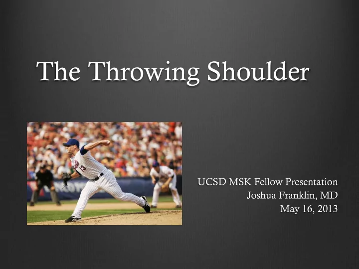
The Throwing Shoulder
UCSD MSK Fellow Presentation Joshua Franklin, MD May 16, 2013
The Throwing Shoulder UCSD MSK Fellow Presentation Joshua Franklin, - - PowerPoint PPT Presentation
The Throwing Shoulder UCSD MSK Fellow Presentation Joshua Franklin, MD May 16, 2013 Objectives Throwing Motion Dead Arm Posterosuperior Impingment GIRD Pathologic Cascade Humeral Retroversion Bennett Lesion Anterosuperior Impingement
UCSD MSK Fellow Presentation Joshua Franklin, MD May 16, 2013
Throwing Motion Dead Arm
Posterosuperior Impingment GIRD – Pathologic Cascade
Humeral Retroversion Bennett Lesion Anterosuperior Impingement Little Leaguer’s Shoulder
6 phases (Fleisig, et al. 1996) 1) Windup 2) Early cocking / Stride 3) Late cocking 4) Acceleration 5) Deceleration 6) Follow through
Elevation of lead leg to highest point Separation of throwing and glove hands
Seroyer et al. 1999
Begins at lead leg max height and ends at stride foot contact Early shoulder abduction and external rotation
Seroyer et al. 1999
Begins with foot contact Ends with maximal abduction and external rotation of the shoulder
Seroyer et al. 1999
Between maximum external rotation and ball release Rapid horizontal adduction and internal rotation of the humerus
Internal rotation velocities up to 7000o/sec (Fleisig et al 1994) Possibly fastest motion in all of sport
Seroyer et al. 1999
Most violent phase Between ball release and maximum humeral internal rotation
Arm outstetched towards home plate
Biceps and brachialis
Decelerating elbow extension
Large distraction forces on posterior soft tissue structures of the glenohumeral joint
Up to 80% of body weight
Seroyer et al. 1999
Between maximum adduction and internal rotation and arm coming to rest Ends with pitcher in the fielding position
Seroyer et al. 1999
Pitch velocity
Proportional to internal rotation velocity of the humerus during acceleration phase
Increased maximum external rotation during late cocking phase
Increased distance for accelerating forces to act
“The slot”
Proprioceptive sense of the external rotation set point needed to obtain maximum velocity
°
fi fi fi
Soft tissue and osseous adaptations allow increased external rotation in late cocking Evenutally some of these adaptations may lead to pathology and “dead arm”
“Any pathologic shoulder condition in which the thrower is unable to throw with preinjury velocity and control because
shoulder.” (Burkhart et al 2003)
Discomfort typically late cocking/early acceleration phase Sudden sharp pain and arm “goes dead”
Mysterious etiology
Psychopathology, posterior glenoid calcs, acromial osteophytes, CA ligament impingement, rotator cuff, biceps tendon, AC joint, microinstability, internal impingement, SLAP
Walch et al first described impingement of undersurface of posterosuperior rotator cuff between greater tuberosity and posterosuperior glenoid and labrum with ABER. Contact can be physiologic in ABER Spectrum of pathologic findings
Undersurface tears of the posterior supraspinatus and anterior infraspinatus where impingement occurs Posterosuperior labral tears Cystic changes and sclerosis posterior greater tuberosity and posterosuperior humeral head and posterior glenoid
’ “ ” fi fi °
Burkhart 2003
Jobe applied this concept to throwing shoulder
Repetitive ABER in late cocking phase Proposed stretching of anterior capsuloligamentous structures in throwers leads to progression
Halbrecht et al 1999
Halbrecht et al. 1999
10 asymptomatic college baseball players Bilateral shoulder MR arthrograms Contact between cuff undersurface and posterosuperior labrum in ABER
Throwing and non-throwing shoulders Likely physiologic
Throwing shoulders only
3 posterosuperior labral tears 2 cuff tears and 2 others with tendinosis 2 posterosuperior humeral head and posterior glenoid cystic changes
No correlation with anterior instability Halbrecht 1999
Giaroli et al, AJR 2005
6 patients surgically confirmed PSI
4 baseball, 1 tennis, 1 swimmer
15 control patients 100% cases
Abnormal PS labrum vs 13% controls Abnormal cuff undersurface vs 27% Cyst like changes in humeral head vs 27%
Cyst like changes
More posterior than typically seen with cuff pathology
22 year old professional pitcher with stiffness and normal pitching velocity
Courtesy of Brady Huang, MD
16 year old female swimmer
Courtesy of Brady Huang, MD
Glenoid internal rotation deficit
Loss in degrees of glenohumeral internal rotation compared with the non-throwing shoulder GIRD in symptomatic shoulders generally > 25o Loss of internal rotation far exceeds gain in external rotation
Caused by posteroinferior capsular contracture Burkhart favors GIRD as initiating a cascade eventually leading to SLAP lesions and dead arm
° ° ° fi ° ° fi fi fi ° ° fi ° ° ° fi ° ° fi ° “ ”
’ ° ° fl ° ’ ’
Burkhart et al 2003
Clinical evidence
60% of 39 professional pitchers with at least 35o GIRD developed shoulder problems requiring them to stop pitching (Verna 1991) Morgan treated 124 pitchers with arthroscopically proven SLAP 2 lesions – all had severe GIRD Kibler found severe GIRD in all 38 overhead athletes treated for proven Type 2 SLAP Donley and Cooper found asymptomatic ptichers only 13o GIRD preseason and 16o GIRD posteseason Kibler found decreased GIRD and 38% decrease in shoulder injuries in a group of tennis players who performed daily posterior capsular stretching compared with control group
PSI normal phenomenon and is not etiology of dead arm
Posteroinferior capsular contracture – GIRD Shift glenohumeral contact point in ABER Hyper-external rotation in ABER Increased shear forces on the biceps anchor and posterosuperior labrum
Peel back mechanism Type II SLAP tear
Increased shear and torsional stress on posterosuperior cuff
Undersurface rotator cuff tears
Burkhart et al 2003
Superior Labrum anterior to posterior Snyder Classification
Type I: Fraying Type II: Tear of biceps labral complex Type III: Bucket handle tear Type IV: Bucket handle tear with extension to Biceps
Morgan Type II subtypes
IIA: anterior extension IIB: posterior extension IIC: anterior and posterior extension
fi fi fi fi “ ” ° °
Burkhart et al 2003
Large distraction forces on posteroinferior capsule during deceleration (750N, 80% BW) Repetitive tensile loading leads to posteroinferior capsular hypertrophy GIRD results
fi fi fi fi fi ’
’
Burkhart et al 2003
PIGHL shifts under humeral head during ABER Contracted PIGHL exerts posterosuperior force on humeral head Posterosuperior shift of the GH contact point
Burkhart et al 2003
ˆ “ ” “CIRCLE CONCEPT” fi fi fi
Contracted PIGHL AIGHL MGHL SGHL Biceps
ANTERIOR POSTERIOR
Courtesy of Brady Huang, MD
Contact point shift allows hyperexternal rotation via 2 mechanisms Increased clearance of the greater tuberosity before internal impingement
Greater arc of external rotation
Decreased CAM effect of humeral head and proximal humeral calcar on anterior capsule
Relative redundancy of anterinferior capsule
° ° fi fi
fi
Burkhart et al 2003
Twisting of biceps tendon with hyperexternal rotation Shearing force directed to posterosuperior labrum Type II SLAP tears
Predominantly Type IIb or IIc
Peel back
Biceps root will shift medial to supraglenoid tubercle
This is likely cause of “dead arm”
fi fi fi fi fi fi
Burkhart et al 2003
Anterior instability reported in dead arm
Positive drive through sign at arthroscopy Scope driven from top to bottom of GH joint without resistance
Burkhart suggests this is due to pseudolaxity not true instability Decreased CAM effect – capsular redundancy Circle concept
Break in labral ring (from SLAP tear) allows channeling of apparent laxity to opposite side of ring where there is disruption.
ˆ “ ” “CIRCLE CONCEPT” fi fi fi
Burkhart et al 2003
Andrews et al (1985) initially proposed deceleration mechanism
High tensile load on long head biceps tendon with deceleration
Cadaver model (Kuhn et al) supports acceleration model
Type 2 SLAP in 90% of specimens from loading biceps in ABER Type 2 SLAP in only 20% of specimens loaded in follow-through 20% less force needed in ABER
Most thrower’s recall late cocking/early acceleration phase as position of injury (Burkhart 2003)
Hypertwist from hyperexternal rotation Torsional and shear overload of cuff undersurface leading to tears
°
fi fi fi
Burkhart et al 2003
Anterior instability
Not part of the inciting pathology of dead arm Anterior capsular failure may be tertiary problem Increased tensile stress on AIGHL with repetitive hyperexternal rotation
Posterosuperior impingement
Not primary pathology in dead arm May be seen in older elite thowers
Hyperxternal rotation in late cocking in excess of 130o Burkhart et al 2003
’ fi ° fi “ ”
⁄ ’ ’ ° °
Most respond to posterior capsular stretching SLAP repair
Morgan and Burkhart report 87% return to preinjury level of performance and velocity
Selective posterior capsulotomy in stretch non-responders
Burkhart et al 2003
Saleem et al AJR 2008 – Pictoral essay
Usefullness of the Abduction and external rotation views in shoulder MR arthrography
ABER view may recreate decentering of humeral head with GIRD
Sanders, Zlatkin, and Montgomery, 2010
Imaging of Glenohumeral Instability (review)
18 year old baseball pitcher with GIRD
Marked thickening of the posterior capsule
Tehranzadeh, Fronek, and Resnick (Clinical Imaging, 2007)
Retrospective study of MR arthrograms in 6 professional pitchers with symptomatic GIRD 5 with arthroscopy
All with undersurface rotator cuff tears 4 SLAP tears, 2 posterior labral fraying Subjective posteroinferior capsular thickening
No standard at the time for measurement
Tuite et al – Skeletal Radiology 2007
26 overhead athletes with GIRD and internal impingement 26 asymptomatic controls
Attempt to diagnose and quantify posterior capsular thickening
Labral length at 8 o’clock – subchondral bone to labral tip Posterior recess angle Thick - capsule labral length
If capsule inserted near labral tip Subchondral bone to where capsule “thinned out to become 1-2mm thick”
All differences statistically significant Limitations
PIGHL difficult to separate from capsule near labral attachment
Labral measurement used as surrogate
Glenoid shape differences not controlled for
i.e. hypoplasia with thick posterior labrum
Somewhat arbitrary thick capsule-labral length end point Developmental variations in capsular attachment not accounted for
Park et al, AJR 2000 showed variable posterior capsular attachment 60% type 1 – directly onto labrum 31% type 2 – junction of labrum and glenoid 9% type 3 – Medial to glenoid
Overlap of measurements Technical differences – Patient positioning, amount of contrast Overhead athletic participation as children not controlled for Labral Length (mm)
Thick capsule – labral length (mm)
Posterior recess (o)
GIRD 6.4 (2.9- 12.5) 8.8 (2.9-16.5) 94 (18-160) Controls 4.9 (2.3-9.5) 5.4 (2.3-11.7) 65 (28-148)
Borrero et al, Skeletal Radiology 2010
34 patients surgically confirmed labral tear with peel back in ABER 29 controls with no peel back
Attempt to describe MR appearance and determine reliability of MR for prospective diagnosis of posterosuperior labral peel back
Position of posterosuperior labrum with respect to glenoid on ABER
Burkhart et al 2003
Posterosuperior labrum = “Reverse C” shape Three point grading system
0 - Apex of reverse C clearly lateral and craniad to glenoid tangent line 1 - Apex flush with glenoid tangent line 2 – Apex clearly medial and caudal to glenoid tangent line on at lease 1 image Grade 0 Grade 2 Grade 1
Grade 0 and Grade 1 considered negative Grade 2 considered peel back 2 blinded readers: excellent inter-rater agreement – Kappa coefficient of 0.9 Sensitivity 73%, Specificity 100%, PPV 100%, NPV 78%
Grade 0 Grade 2 Grade 1
Additional analysis of value of ABER for diagnosis of SLAP 5 of 34 patients
SLAP tear only evident on ABER No labral tear evident on standard sequences
Posteromedial angle between the axis of the elbow and the axis through the center of the humeral head Normal values range from 10o-40o Increased retroversion
Seen in dominant arms of overhead athletes Allows increased external rotation before humeral head contrained by anterior capsule
Hernigou 2002 Reagan et al. 2002
Pieper, AJSM 1998
51 professional handball players
Retrotorsion angle average of 9.4o greater on dominant side No side to side difference in retroversion of controls
Players with chronic shoulder pain - no side to side difference Increased retroversion adaptive response allowing more external rotation before excessive strain on shoulder soft tissues
Crockett et al, AJSM 2002
25 professional pitchers vs 25 controls Retroversion in dominant arm of pitchers – 40o
Significantly greater than non dominant arm (23o) Significantly greater than dominant arm of nonthrowers (18o)
Nonthrowers
No significant difference between dominant (18o) and nondominant (19o)
Reagan et al, AJSM 2002
54 asymptomatic college baseball players Significantly greater retroversion in dominant arm
Dominant arm – 36.6o Nondominant arm – 26o
Multiple different techniques
Radiographic techniques complicated CT preferred method
CT Reference lines
Line perpendicular to the proximal articular surface of the humeral head Distal reference Line
Transepicondylar line Trochlear tangent line Forearm axis Reagan et al. 2002 Hernigou 2002
Ossification near the posteroinferior glenoid in
Bennett (1941) originally believed traction injury at attachment of the long head
Generally accepted as traction injury of posteroinferior capsule and PIGHL
Shah and Tung 2009
Best evaluated by CT and radiographs Extra-articular crescentic calcification/ossification near the glenoid attachment of PIGHL Special radiographic views may be helpful
Bennett or modified Bennett view
ABER with beam angled 5o cephalad
Stryker notch view
Hand on head with elbow straight up Beam angled 10o cephalad Wright and Paletta 2004 Modified Bennett Stryker Notch View
http://depts.washington.edu/shoulder/X-Rays.htm
Meister et al 1999
Low signal abnormality within capsule May contain high T1 signal from marrow Possibly associated with capsular thickening Pericapsular edema
More acute injury?
Sanders et al 2010
Chung CB and Steinbach LS. MRI of the upper extremity: elbow, wrist, and hand. 2009. (Anecdotal data per Dr. Chung)
Controversial clinical significance and treatment Associated with posterior shoulder pain
ABER position – late cocking/early acceleration Adduction/internal rotation – follow through/deceleration
Frequently seen in asymptomatic overhead athletes
12 of 55 (22%) asymptomatic MLB pitchers (Wright et al 2004)
Frequently associated with other intra-articular pathology
Posterior and posterosuperior labral tears Undersurface rotator cuff tears
Variable results for surgical excision
Lombardo et al, AJSM 1977
3 pitchers with open excision of exostosis and posterior labral debridement All 3 returned to pre-injury level of performance
Ferrari et al, AJSM 1994
7 baseball players with shoulder pain and Bennett’s lesion 6 labral tears and 6 undersurface rotator cuff abnoramlities repaired Bennett lesion not treated 6/7 returned to preinjury level
Ferrari et al, 1994
Meister et al, AJSM 1999
22 overhead athletes with shoulder pain and Bennett’s 11 patients - exostosis debrided 21 patients - undersurface cuff tears debrided 20 patients - Labral “fraying” debrided
15 posterior, 4 anterior, 1 superior
10 of 18 patients with follow up returned to preinjury level > than 1 year
Exostosis debridement no effect on return
Still recommended excision of large exostosis in patients with posterior shoulder pain
Yoneda, et al 2002
16 baseball players with arthroscopic resection of symptomatic Bennett’s
Posterior shoulder pain with throwing Pain with throwing reduced by lido injection into Bennett’s Posteroinferior GH joint tenderness
11/16 returned to preinjury level of performance Many associated abnormalities also treated
6 articular sided cuff tears debrided 4 posteroinferior labral tears repaired 4 “biceps and labral complex injuries” repaired or debrided
Both involve PIGHL PIGHL thickening sometimes seen with Bennett lesion Tuite et al, Skeletal Radiology 2007
Different response to same repetitive injury ? Spectrum of injury where some with GIRD go on to Bennett’s?
Meister et al, AJSM 2002
“An asymmetric loss of internal rotation and increased external rotation was noted in all 22 patients”
Shah and Tung 2009 Sanders et al, 2010
Impingment of undersurface of biceps pulley and subscapularis tendon against anterosuperior glenoid rim with adduction and internal rotation Gerber and Sebesta, 2000
16 pts c/o pain with anterior elevation and internal rotation 12/16 in overhead professions (masonry) 3 isolated pulley lesions 10 pulley lesions + undersurface tears of upper fibers of subscap 3 intact pulley with undersurface subscap 16/16 impingment with horizontal adduction and internal rotation at arthroscopy Likely from repetitive overhead movements
http://mlbblogger.files.wordpress.com/2013/03/maestri-follow-through.jpg?w=555&h=312
Habermeyer et al 2004
89 Patients with pulley lesions 4 groups (% with ASI at arthroscopy)
Role of subscap tears
Increased ASI with Subscap involvement LHB loses stabilizing effect on GH joint Anterosuperior humeral head translation Increased ASI
Pulley lesion
Degenerative Traumatic – forcefully stopped overhead throwing motion
Very little in radiology literature Barile et al 2013
23 overhead athletes with suspected pulley lesions Excellent correlation with Arthroscopy
2 downgraded from I to normal 2 upgraded from III to IV
Increased ASI with subscap involvement MR less accurate for AS Labrum
Type 4: 6 MR vs 10 Arthro Type 3: 2 MR vs 4 Arthro Habermeyer class I II III IV MR 3 5 7 8 Arthroscopy 2 1 5 5 10 ASI - Arthroscopy 2 10
Radiol med (2013) 118:112–122
fibr all’artr all’artr fibr All’artr all’artr classificati è fi subacromion–deltoid fiv confirmed fibres; confirmed;
a b c d
’imma ’imma ’esame Radiol med (2013) 118:112–122
’IAS è [1–3] identificazione 4–6]. flitto hé dell’intervallo ’intervallo è fino 10–14]. dal LGOS e dal LCO [8, 15–17]. identified “nonoutlet” [1–3] 4–6]. conflicting
a b c d
confirms ’imma
Proximal humeral physis overuse injury
Rotational stress during overhead throwing
Gradual onset of shoulder pain with throwing and tenderness at lateral aspect
Most commonly in early to mid teenage years
Physis vulnerable during period of rapid growth
Mechanism of injury
Injury to metaphyseal vessels supplying physis
Necessary for chondrocyte death and cartilage mineralization Prolonged chondrocyte survival and extension into metahpysis
Fleming et al 2004
Radiographic findings
May be normal if symptoms less than 10 days Proximal humeral physeal widening Less common findings
Metaphyseal demineralization
Fragmentation or cystic changes lateral aspect of the proximal humeral metaphysis Carson and Gasser 1998
Hatem et al, 2007
4 pitchers age 12-14 with MRI for shoulder pain 3/4 widened physis
Possibly related to imaging early after symptom onset in 1 patient (3 weeks)
4/4 metaphyseal bone marrow edema 3/4 epiphyseal marrow edema 2/4 periosteal edema
Obembe et al, 2007
4 adolescent overhead athletes (11-15) Extension of physeal signal into metaphysis Metaphyseal bone marrow edema No epiphyseal abnormalities No periosteal edema
Conservative management Rest / throwing restriction Gradually resume throwing Carson and Gasser, 1998
21/23 patients returned to throwing after average rest of 3 months 2 patients still resting at time of publication Radiographic appearance lags behind clinical response
Carson and Gasser, 1998
The throwing shoulder is an evolving and controversial subject GIRD initiating the pathologic cascade to SLAP tear is the leading current theory to explain “dead arm” Knowledge of injury patterns specific to the throwing shoulder can help the radiologist identify the total spectrum
the treating orthopedic surgeon.
Burkhart SS, Morgan CD, Kibler WB. The disabled throwing shoulder: Spectrum of pathology Part I: Pathoanatomy and biomechanics. Arthroscopy 2003; 19: 404–420. Burkhart SS, Morgan CD, Kibler WB. The disabled Throwing Shoulder: Spectrum of pathology Part II: Evaluation and Treatment of SLAP lesions in throwers. Arthroscopy 2003; 19 :531-539. Burkhart SS, Morgan CD, Kibler WB. The disabled Throwing Shoulder: Spectrum of pathology Part III: The SICK scapula, scapular dyskinesis, the kinetic chain, and rehabilitation. Arthroscopy 2003; 19: 641-661. Barile A, Lanni G, Conti L, Mariani S, Calvisi V , Castagna A, Rossi F, Masciocchi C. Lesions of the biceps pulley as cause of anterosuperior impingement of the shoulder in the athlete: potentials and limits of MR arthrography compared with arthroscopy. Radiol med 2013; 118:112-122. Borrero CG, Casagranda BU, Towers JD, Bradley JP. Magnetic resonance appearance of posterosuperior labral peel back during humeral abduction and external rotation. Skeletal Radiol 2010; 39:19-26 Carson WG, Gasser SI. Little leaguer’s shoulder, a report of 23 cases. Am J Sports Med 1998; 26(4): 575-580. Chang D, Mohana-Borges A, Borso M, Chung CB. SLAP lesions: Anatomy, clinical presentation, MR imaging diagnosis and characterization. Eur J Rad 2008; 68: 72-87. Chung CB, Steinbach LS. MRI of the upper extremity: elbow, wrist, and hand. Lippincott Williams & Wilkins, 2009 Crockett HB, Gross LB, Wilk KE, Schwartz ML, Reed J, O’Mara J, Reilly MT, Dugas JR, Meister K, Lyman S, Andrews JR. Osseous adaptation and range of motion at the glenohumeral joint in professional baseball
Ferrari JD, Ferrari DA, Coumas J, Pappas AM. Posterior ossification of the shoulder: the Bennett Lesion : Etiology, Diagnosis, and Treatment. Am J Sports Med 1994; 22: 171-176 Fleming JL, Hollingsworth CL, Squire DL, Bisset GS. Little leaguer’s shoulder. Skeletal Radiol 2004; 33:352-354. Giaroli EL, Major NM, Higgins LD. MRI of internal impingement of the shoulder. Am J Roentgenol 2005; 185: 925-929. Gerber C, Sebesta A. Impingment of the deep surface of the subscapularis tendon and the reflection pulley on the anterosuperior glenoid rim: A preliminary report. J Shoulder Elbow Surg 2000; 9:483- 490. Habermeyer P, Magosch P, Pritsch M, Scheibel T, Lichtenberg S. Anterosuperior impingement of the shoulder as a result of pulley lesions: a prospective arthroscopic study. J Shoulder Elbow Surg 2004; 13: 5-12. Halbrecht JL. Internal impingement of the shoulder: comparison of findings between the throwing and nonthrowing shoulders of college baseball players. Arthroscopy 1999; 15:253–258. Hatem SF, Recht MP, Profitt B. MRI of little leaguer’s shoulder. Skeletal Radiol 2006; 35:103-106. Hernigou P, Duparc C, Hernigou A. Determining humeral retroversion with computed
Kirchoff C, Imhoff AB. Posterosuperior and anterosuperior impingment of the shoulder in
Leonard J, Hutchinson MR. Shoulder injuries in skeletally immature throwers: review and current thoughts. Br J Sports Med 2010; 44: 306-310. Lyman S, Fleisig GS, Andrews JR, Osinski ED. Effect of pitch type, pitch count, and pitching mechanics on risk of shoulder pain in youth baseball pitchers. Am J Sports Med 2002; 30(4) 463-468. Meister K, Andrews JR, Batts J, et al: Symptomatic thrower’s exostosis. Am J Sports Med 27(2): 133–136, 1999 Miniaci A, Mascia AT, Salonen DC, Becker EJ. Magnetic resonance imaging of the shoulder in asymptomatic professional baseball pitchers. Am J Sports Med 2002; 30(1): 66-73. Obembe OO, Gaskin CM, Taffoni MJ, Anderson MW . Little leaguer’s shoulder (proximal humeral epiphysiolysis): MRI findings in four boys. Pediatr Radiol 2007; 37:885-889. Park YH, Lee JY, Moon SH, Mo JH, Yang BK, Hahn SH, Resnick D. MR arthrography of the labral capsular ligamentous complex in the shoulder: Imaging variations and pitfalls. Pieper HG. Humeral torsion in the throwing arm of handball players. Am J Sports Med 1998; 26(2): 247-253. Reagan KM, Meister K, Horodyski MB, Werner DW , Carruthers C, Wilk K. Humeral retroversion and its relationship to glenohumeral rotation in the shoulder of college baseball players. Am J Sports Med 2002; 30(3): 354-360. Roger B, Skaf A, Hooper AW , Lektrakul N, Yeh LR, Resnick D. Imaging findings in the dominant shoulder
with arthroscopic correlation. Am J Roentgenol 1999; 172:1371-1380. Saleem AM, Lee JK, Novak LM. Usefulness of abduction and external rotation views in shoulder MR
Sanders TG, Zlatkin MD, Montgomery J. Imaging of glenohumeral instability. Semin Roentgenol 2010; 45(3)160-179. Seroyer ST, Nho SJ, Bach BR, Bush-Joseph CA, Nicholson GP, Romeo AA. The kinetic in overhand pitching: Its potential role for performance enhancement and injury prevention. Sports Health 2010; 2: 135-146. Shah N, Tung GA. Imaging signs of posterior glenohumeral instability. Am J Roentgenol 2009; 192:730-735. Song JC, Lazarum ML, Song AP. MRI findings in little leaguer’s shoulder. Skeletal Radiol 2006; 35:107-109. Tehranzadeh AD, Fronek J, Resnick D. Posterior capsular fibrosis in professional baseball pitchers: case series of arthrographic findings in six patients with glenohumeral internal rotational deficit. Clinical Imaging 2007; 31:343-348 Tuite MJ, Peterson BD, Wise SM, Fine JP, Kaplan LD, Orwin JF. Shoulder MR arthrography of the posterior labrocapsular complex in overhead throwers with pathologic internal impingement and internal rotation deficit. Skeletal Radiol 2007; 36:495–502 Whiteley R. Baseball throwing mechanics as they relate to pathology and performance – a review. J Sports Sci Med 2007; 6: 1-20. Wright RW , Paletta GA. Prevalence of the Bennett lesion of the shoulder in major league Pitchers. Am J Sports Med 2004 32: 121 Yoneda M, Nakagawa S, Hayashida K, et al: Arthroscopic removal of symptomatic Bennett lesions in the shoulders of baseball players: Arthroscopic Bennett-plasty. Am J Sports Med 30(5): 728–736, 2002