Unusual Presentation of Flexor Tendon Sheath Massive Ganglion Saleeb - - PDF document

unusual presentation of flexor tendon sheath massive
SMART_READER_LITE
LIVE PREVIEW

Unusual Presentation of Flexor Tendon Sheath Massive Ganglion Saleeb - - PDF document

Journal of Surgery and Operative Care Volume 1 | Issue 1 ISSN: 2455-7617 Open Access Case Report Unusual Presentation of Flexor Tendon Sheath Massive Ganglion Saleeb H *1 and Kanvinde R 2 1 Orthopaedic Registrar, Betsi Cadwaladr University


slide-1
SLIDE 1

Annex Publishers | www.annexpublishers.com Volume 1 | Issue 1

Unusual Presentation of Flexor Tendon Sheath Massive Ganglion

Saleeb H*1 and Kanvinde R2

1Orthopaedic Registrar, Betsi Cadwaladr University Health Board 2Consultant Orthopaedic Surgeon, Betsi Cadwaladr University Health Board

*Corresponding author: Saleeb H, Orthopaedic Registrar, Betsi Cadwaladr University Health Board, E-mail:

mr.hany.saleeb@gmail.com

Case Report Open Access

Abstract

Citation: Saleeb H, Kanvinde R (2016) Unusual Presentation of Flexor Tendon Sheath Massive Ganglion. J

Surg Oper Care 1(1): 105. doi: 10.15744/2455-7617.1.105

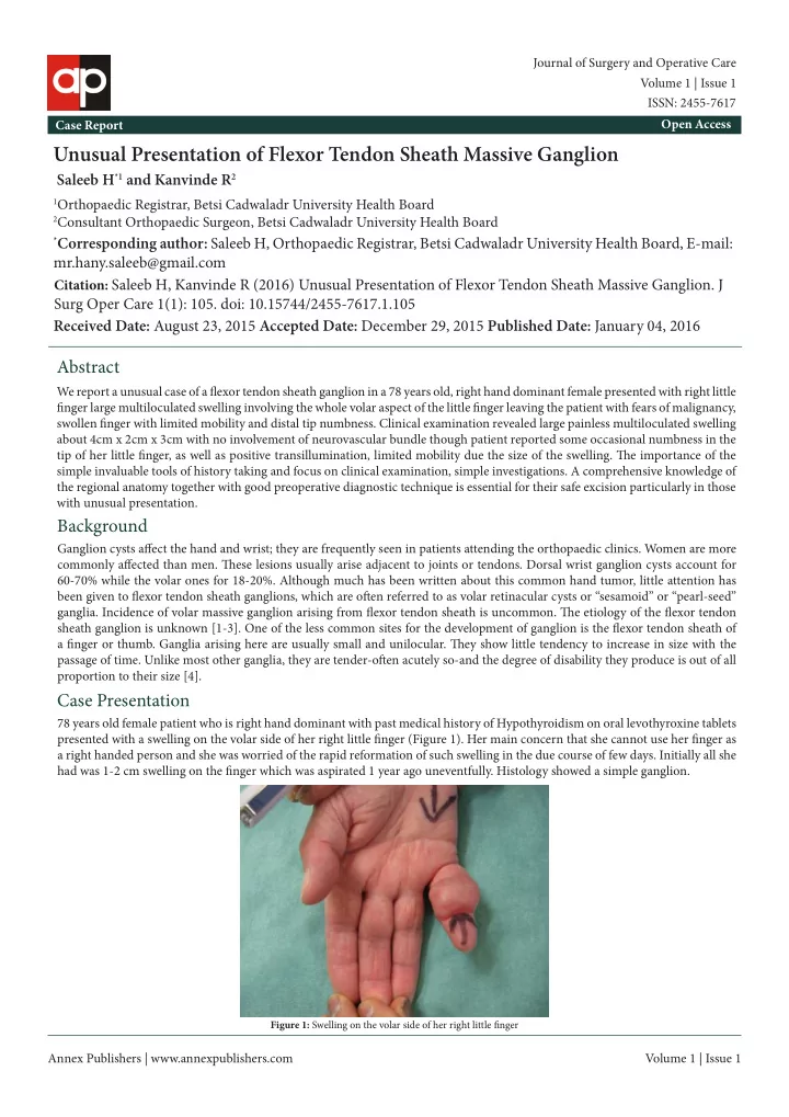
We report a unusual case of a fmexor tendon sheath ganglion in a 78 years old, right hand dominant female presented with right little fjnger large multiloculated swelling involving the whole volar aspect of the little fjnger leaving the patient with fears of malignancy, swollen fjnger with limited mobility and distal tip numbness. Clinical examination revealed large painless multiloculated swelling about 4cm x 2cm x 3cm with no involvement of neurovascular bundle though patient reported some occasional numbness in the tip of her little fjnger, as well as positive transillumination, limited mobility due the size of the swelling. Tie importance of the simple invaluable tools of history taking and focus on clinical examination, simple investigations. A comprehensive knowledge of the regional anatomy together with good preoperative diagnostic technique is essential for their safe excision particularly in those with unusual presentation.

Background

Ganglion cysts afgect the hand and wrist; they are frequently seen in patients attending the orthopaedic clinics. Women are more commonly afgected than men. Tiese lesions usually arise adjacent to joints or tendons. Dorsal wrist ganglion cysts account for 60-70% while the volar ones for 18-20%. Although much has been written about this common hand tumor, little attention has been given to fmexor tendon sheath ganglions, which are ofuen referred to as volar retinacular cysts or “sesamoid” or “pearl-seed”

  • ganglia. Incidence of volar massive ganglion arising from fmexor tendon sheath is uncommon. Tie etiology of the fmexor tendon

sheath ganglion is unknown [1-3]. One of the less common sites for the development of ganglion is the fmexor tendon sheath of a fjnger or thumb. Ganglia arising here are usually small and unilocular. Tiey show little tendency to increase in size with the passage of time. Unlike most other ganglia, they are tender-ofuen acutely so-and the degree of disability they produce is out of all proportion to their size [4].

Case Presentation

78 years old female patient who is right hand dominant with past medical history of Hypothyroidism on oral levothyroxine tablets presented with a swelling on the volar side of her right little fjnger (Figure 1). Her main concern that she cannot use her fjnger as a right handed person and she was worried of the rapid reformation of such swelling in the due course of few days. Initially all she had was 1-2 cm swelling on the fjnger which was aspirated 1 year ago uneventfully. Histology showed a simple ganglion.

Figure 1: Swelling on the volar side of her right little fjnger

Received Date: August 23, 2015 Accepted Date: December 29, 2015 Published Date: January 04, 2016 Volume 1 | Issue 1

Journal of Surgery and Operative Care ISSN: 2455-7617

slide-2
SLIDE 2

Annex Publishers | www.annexpublishers.com Volume 1 | Issue 1 Journal of Surgery and Operative Care 2 Since then she started noticing rapid formation a swelling almost involving the whole fjnger leaving her with nonfunctional fjnger with numbness on its tip. Clinical examination revealed large painless multiloculated swelling about 4cm x 2cm x 3cm with no involvement of neurovascular bundle though patient reported some occasional numbness in the tip of her little fjnger. Tie swelling was not mobile with attempts to bend fjngertip with limited fjnger mobility due the size of the swelling especially at PIP joint. Positive transillumination test (Figure 2).

Investigations

Giant Cell Tumor of the Tendon Sheath

Giant cell tumors of the tendon sheath are the second most common tumors of the hand, with simple ganglion cysts being the most common. Chassaignac fjrst described these benign sofu-tissue masses in 1852, and he overstated their biologic potential in referring to them as cancers of the tendon sheath. Giant cell tumors of the tendon sheath (GCTTS) are uncommon and usually benign lesions that arise from the tendon sheath. It is unclear whether these lesions represent neoplasms or simply reactive masses. It is also known as pigmented villonodular tumor of the tendon sheath (PVNTS) or extra-articular pigmented villonodular tumor

  • f the tendon sheath [5,6]. Tie clinical difgerential diagnosis may include foreign body granuloma, necrobiotic granuloma,

tendinous xanthoma, fjbroma of the tendon sheath, infection, ganglion cyst, rheumatoid nodule, epidermoid cyst, lipoma, and a knuckle pad, among other less common entities (Figure 3 and 4). Many of these entities can ofuen be excluded with careful history taking and physical examination [7,8].

Figure 2: Transillumination test

X rays did not show any bony abnormalities. Ultrasound scan showed multiloculated swelling related to the tendon.

Difgerential Diagnosis

Figure 3: Intraoperative images 1

slide-3
SLIDE 3

Annex Publishers | www.annexpublishers.com Volume 1 | Issue 1 Journal of Surgery and Operative Care 3 Tiough a variety treatment modalities had been advocated for ganglions in general. In our case surgical excision afuer careful preoperative planning as shown in the fjgures taking in consideration the skin condition, size of the defect was the best option (Figure 5). Other modalities of treatment as aspiration of the cyst followed by injection usually with steroids was not an option afuer recurrence.

Figure 4: Intraoperative size 4cmx3cmx2cm

Treatment

Figure 5: Afuer surgical excision

Patient postoperatively had functional fjnger with intact neurovascular bundle.

Outcome and Follow-Up

Tiere are a few reported cases of fmexor tendon sheath ganglion reported. Our search showed few reports in the literature on the results of surgery for ganglia of the fmexor tendon sheaths of the digits [5,3]. Case reports on massive fmexor tendon sheath ganglions were not abundant. Our search showed only one case of a fmexor tendon sheath ganglion in a 17-year old female who presented with pain, triggering and a swelling at the base of her right ring fjnger. Ganglion was measuring only 0.5×0.8×0.4 cm in size was removed from the A2 pulley area. [9].

Discussion

Unusual presentations of ganglions may be encountered inspite of the atypical age, size and extension of the swelling. Tie diagnosis may be established preoperatively by History taking and physical examination and non-expensive investigations as x rays and

  • ultrasound. Planning the surgical incision in such cases can ensure safe dissection and complete excision. Etiology of ganglions

remain not very well understood. Tie usual size is about 1-2 cm, but some atypical cysts exist. Tie cyst presents frequently as painless, small sofu tissue mass. A comprehensive knowledge of the regional anatomy together with good preoperative diagnostic technique is essential for their safe excision particularly in those with unusual presentation.

Learning Points/Take Home Messages

slide-4
SLIDE 4

Journal of Surgery and Operative Care 4 Annex Publishers | www.annexpublishers.com Volume 1 | Issue 1

References

  • 1. Angelides AC (1999) Ganglions of the hand and wrist. Operative hand surgery 2171-83.
  • 2. Carp L, Stout AP (1928) A study of ganglion with special reference to treatment. Surg Gynecol Obstet 46: 460-8.

Submit your next manuscript to Annex Publishers and benefjt from:

Submit your manuscript at http://www.annexpublishers.com/paper-submission.php → Easy online submission process → Rapid peer review process → Open access: articles available free online → Online article availability soon afuer acceptance for Publication → Better discount on subsequent article submission → More accessibility of the articles to the readers/researchers within the fjeld

  • 3. Matthews P (1973) Ganglia of the fmexor tendon sheaths of the hand. J Bone Joint Surg 55: 612-7.
  • 4. Herrington JL, Edwards LW (1955) Ganglion cysts arising in unusual locations. Ann Surg 142: 900-3.
  • 5. Finsen V, Håberg Ø, Borchgrevink GE (2013) Surgery for ganglia of the fmexor tendon sheath. Orthop Rev 5: e6.
  • 6. Messoudi A, Fnini S, Labsaili N, Ghrib S, Rafai M, et al. (2007) Giant cell tumors of the tendon sheath of the hand: 32 cases. Chir Main 26: 165-9.
  • 7. Sluijmer HC, Becker SJ, Ring DC (2013) Benign upper extremity tumors: factors associated with operative treatment. Hand (N Y) 8: 274-81.
  • 8. Mulkeen D, Chin-Aleong J, Callaghan J, McCann J, Regan PJ (2000) Fibrous histiocytoma of tendon sheath of the hand. Ir Med J 93: 236-9.
  • 9. Gunaseelan P, Jeremy P, Chua C, Rashdeen F (2015) Ganglion of the Flexor Tendon Sheath at the A2 Pulley. Malay Ortho J 9: 28-9.