
SLIDE 1
Jaan Männik
Saturday Morning Physics Lecture
- Mar. 11, 2017
V B m V B m m m m T k F
s c B tot
- ln
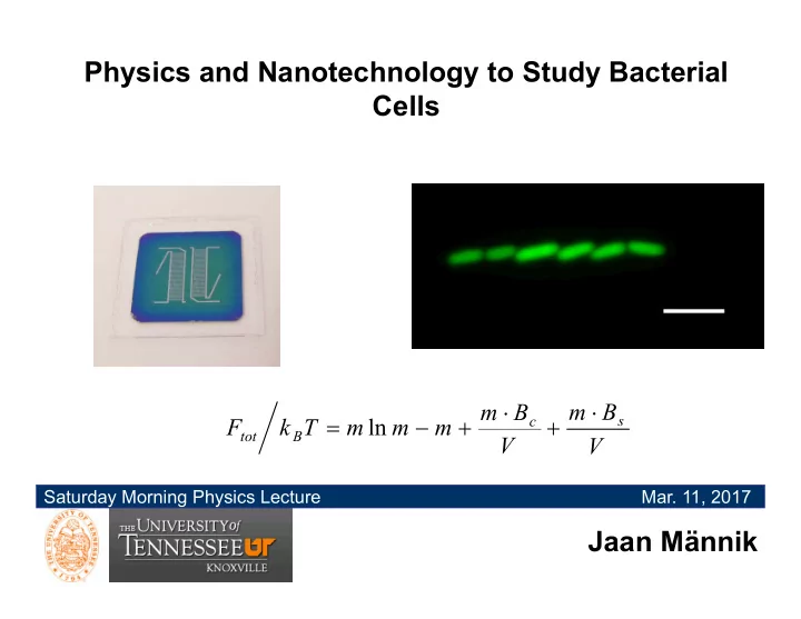
Physics and Nanotechnology to Study Bacterial Cells m B m B - - PowerPoint PPT Presentation
Physics and Nanotechnology to Study Bacterial Cells m B m B s c F k T m ln m m tot B V V Saturday Morning Physics Lecture Mar. 11, 2017 Jaan Mnnik Impact of physics on studies of living systems
Saturday Morning Physics Lecture
V B m V B m m m m T k F
s c B tot
The standard microbiology tools are not suitable to follow:
An agar plate Cell culture tubes
2 m
Device to extract bacterial DNA
Science 301 (2003) 188
Science 305 (2004) 1622
PNAS 104 (2007) 11889
Sequencing Antibiotic resistance studies Ecology, evolution Bacteria and tissue interactions
D.Huh et al. Science 328 (2010) 1622
fluorescent microscopy
100 m
shaped channels.
Use electron beam or photolithography to write pattern of channels Develop resist Reactive ion etch Si wafer Lift-off resist Close channels Repeat the process for different channel heights
PMMA Si
Si PDMS coated glass coverslip electron beam channels
Drill access holes
1 µm
to using a RIE cryoetch process
1 µm 1 µm
D
cytoplasm cytoplasm
Escherichia coli Bacillus subtilis
1 µm
1 µm
D
1 µm
D
molecular level they are different than humans are from roundworms.
Wikipedia
1 µm
electrical motor
inner membrane
cascade) control direction of rotations
channel chamber chamber
0.5 1.0 1.5 2.0 2.5 10 20 30
<v> [m/s] W [m]
motile in channels which are only 30-40% wider than their diameter
their ability to swim but ..
channel chamber chamber
W = 0.6 m
5 hrs 49 hrs 0 hrs
10 µm
population of aberrantly shaped bacteria
which widths are smaller than their diameters
inflated balloon)
higher osmotic pressure can bacterium maintain in its interior
channel wall channel wall
cell wall 3 nm cell wall 30-40 nm
Step # pmax = 2 atm
pmax
pmax = 3 atm
where cell behavior can be studied using high resolution optical microscope and bioanalytical tools.
despite their long flagella. Both species retain ability to swim in channels which only 30% exceed their body diameter
that they use growth and division.
they can penetrate.
Jacque Monod, “What is Valid for E. coli is also valid for the elephant”
FtsZ MatP tetramer SlmA tetramer
Wikipedia
1 μ
HupA-mCherry labelled chromosome
1 μ
HupA-mCherry labelled chromosome
1 μ
chromosome (nucleoid) Z-ring constriction
Go to the right place !!!
PDB: 1FSz
PDB: 1FSz
1 2
1 2
x [m] y [m]
Min operon: minC, minD, minE
N.K. Tonhat et al EMBO J. 30 (2011) 154
WT slmA minC minC
slmA
Evidence for divisome localization mechanisms independent of the Min system and SlmA in Escherichia coli, M. W. Bailey, P. Bisicchia, B. T. Warren, D. J. Sherratt and J. Männik, PLoS
2 0.0 0.5 1.0
Intensity (arb. units) Length along the long axes [m]
Xz Xn
phase DAPI GFP
0.0 0.2 0.4
0.0 0.2 0.4
Xz/L
Xn/L slmA min
0.00 0.15 0.30 20 40 60 counts Xz-Xn [m]
slmA min
= 66 nm
slmA min
time [min] Length along long axes [m]
Z-ring Chromosome
Length along long axes [m]
slmA min 2 m
2 4 6 1 2
I [arb. units] Length along long axes [m]
nucleoid Z-ring
ΔslmA ΔminC ΔzapA ΔslmA ΔminC ΔzapB
Misplaced Misplaced
slmA min matP
0.0 0.5 1.0 20 40 60 counts Xz-Xn [m] N=123
0.0 0.5 1.0 20 40 60 80 counts Xz-Xn [m] N=218
0.0 0.5 1.0 20 40 60
counts Xz-Xn [m]
N=145
= 202 nm = 150 nm = 222 nm
for positioning of bacterial Z-ring via a negative regulation.
in mid-cell as a result of entropic force. This spontaneous process appears ultimately to control the placement of cell division proteins.
have found a positive regulatory mechanism that promotes Z-ring formation at the vicinity of the Ter region of the chromosome.
Matthew Bailey, UTK George Siopsis, UTK Dan Castillo, UTK Jaana Männik, UTK Paola Bisicchia, Oxford Univ. David Sherratt, Oxford Univ. Piet de Boer, Case Western Reserve Univ. Alex Dajkovic, Univ. of Paris 5 Rodrigo Lamothe-Reyes, McGill Univ. Cees Dekker, Delft University of Tech. Conrad Woldringh, Univ. of Amsterdam Arieh Zaritsky, Ben Gurion University NSF CAREER award