Basic Structure of a Blood Vessel The wall of a blood vessel consists of three layers, or tunics, of different tissues: 1-An epithelial inner lining THE TUNICA INTERNA (INTIMA) 2-A middle layer of smooth muscles TUNICA MEDIA 3- A connective tissue outer covering. TUNICA EXTERNA (ADVENTIA)
The wall of a blood vessel consists of three layers, or tunics, of - - PowerPoint PPT Presentation
Basic Structure of a Blood Vessel The wall of a blood vessel consists of three layers, or tunics, of different tissues: 1-An epithelial inner lining THE TUNICA INTERNA (INTIMA) 2-A middle layer of smooth muscles TUNICA MEDIA 3- A connective
1- 2- 3-
1-Tunica Interna (Intima)
The tunica interna forms the inner lining of a blood vessel and is in direct contact with the blood as it flows through the lumen The main component of the intima is simple sqaumous epithelium Called
ENDOTHELIUM
that lines the inner surface of the entire cardiovascular system (heart and blood vessels)
2-Tunica Media
The tunica media (media middle) is made of a muscular (smooth muscle) and connective tissue layer smooth muscle cells, which extend circularly around the lumen like a ring
3-Tunica Externa
The outer covering of a blood vessel, the tunica externa (outermost)
consists of elastic and collagen fibers
The tunica externa contains numerous nerves and tiny blood vessels that supply the tissue of the vessel wall. `
An increase in
sympathetic stimulation
typically stimulates the smooth muscle to contract and narrowing the lumen. Such a decrease in the diameter of the lumen of a blood vessel is called
vasoconstriction
In contrast, decreases, or in the presence of certain chemicals (such as nitric oxide) or in response to blood pressure, smooth muscle fibers relax. The resulting increase in lumen diameter is called
vasodilatation
!
STRUCTURE AND FUNCTION OF BLOOD VESSELS
The five main types of blood vessels are
1- ARTERIES 2-ARTERIOLES 3-CAPILLARIES 4-VENULES 5-VEINS
B-ARTERIOLES As the arterioles enter a tissue, they branch into numerous tiny : vessels called D-VENULES These in turn merge to form progressively larger blood vessels called veins. 1-ARTERIES carry blood away from the heart to other organs A-LARGE, ELASTIC ARTERIES leave the heart and divide into B-MEDIUM-SIZED, MUSCULAR ARTERIES that branch out into the various regions of the body. Medium-sized arteries then divide into small arteries, which in turn divide into still smaller arteries called : C-CAPILLARIES The thin walls of capillaries allow the exchange of substances between the blood and body tissues. Groups of capillaries within a tissue reunite to form small veins called: E-Veins are the blood vessels that convey blood from the tissues back to the heart:
Small arteries
arterioles are abundant microscopic vessels that regulate the flow of blood into the capillary. Arterioles have a thin tunica interna
2-Arterioles
resistance vessels
3-Capillaries
The smallest of blood vessels, connect the arterial outflow to the venous return The flow of blood from a metarteriole through capillaries and into a postcapillary venule (venule that receives blood from a capillary) is called the microcirculationof the body. The primary function of capillaries is the exchange of substances between the blood and different tissues
The body contains three different types of capillaries:
A- Continuous Capillaries
B-Fenestrated Capillaries
C-sinusoids
Most capillaries are a-CONTINUOUS CAPILLARIES, in which the plasma membranes of endothelial cells form a continuous tube inside the capillary Continuous capillaries are found in the central nervous system, lungs, skin, muscle tissue, and the skin
B-fenestrated capillaries The plasma membranes of the endothelial cells in these capillaries have many fenestrations. Can be found, for example, in kidneys, villi of the small intestine
C-Sinusoids are wider and more winding than other capillaries. Their endothelial cells may have unusually large fenestrations. For example, newly formed blood cells enter the bloodstream through the sinusoids of red bone marrow. Can be found, for example, in The spleen, the liver
4-Venules They have thin walls Venules drain the capillary blood and begin the return flow
- f blood back toward the heart
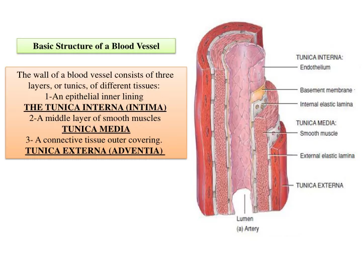
Although veins are composed of essentially the same three layers as arteries, the relative thicknesses of the layers are different.
- The tunica interna of veins is thinner than that of
arteries;
- the tunica media of veins is much thinner than
in arteries, with relatively little smooth muscle and elastic fibers.
- The tunica externa of veins is the thickest layer
and consists of collagen and elastic fibers.
5-Veins
Many veins, especially those in the limbs, also contain valves, thin folds of tunica The valves aid in venous return by preventing the backflow of blood Vein